Chlorine »
PDB 5c2e-5caj »
5c6b »
Chlorine in PDB 5c6b: Crystal Structure of Prefusion-Stabilized Rsv F Variant Sc-Tm
Protein crystallography data
The structure of Crystal Structure of Prefusion-Stabilized Rsv F Variant Sc-Tm, PDB code: 5c6b
was solved by
J.S.Mclellan,
J.P.M.Langedijk,
with X-Ray Crystallography technique. A brief refinement statistics is given in the table below:
| Resolution Low / High (Å) | 44.95 / 2.40 |
| Space group | P 41 3 2 |
| Cell size a, b, c (Å), α, β, γ (°) | 168.200, 168.200, 168.200, 90.00, 90.00, 90.00 |
| R / Rfree (%) | 17.8 / 21.1 |
Chlorine Binding Sites:
The binding sites of Chlorine atom in the Crystal Structure of Prefusion-Stabilized Rsv F Variant Sc-Tm
(pdb code 5c6b). This binding sites where shown within
5.0 Angstroms radius around Chlorine atom.
In total 2 binding sites of Chlorine where determined in the Crystal Structure of Prefusion-Stabilized Rsv F Variant Sc-Tm, PDB code: 5c6b:
Jump to Chlorine binding site number: 1; 2;
In total 2 binding sites of Chlorine where determined in the Crystal Structure of Prefusion-Stabilized Rsv F Variant Sc-Tm, PDB code: 5c6b:
Jump to Chlorine binding site number: 1; 2;
Chlorine binding site 1 out of 2 in 5c6b
Go back to
Chlorine binding site 1 out
of 2 in the Crystal Structure of Prefusion-Stabilized Rsv F Variant Sc-Tm
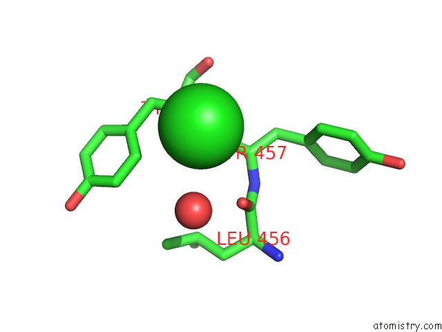
Mono view
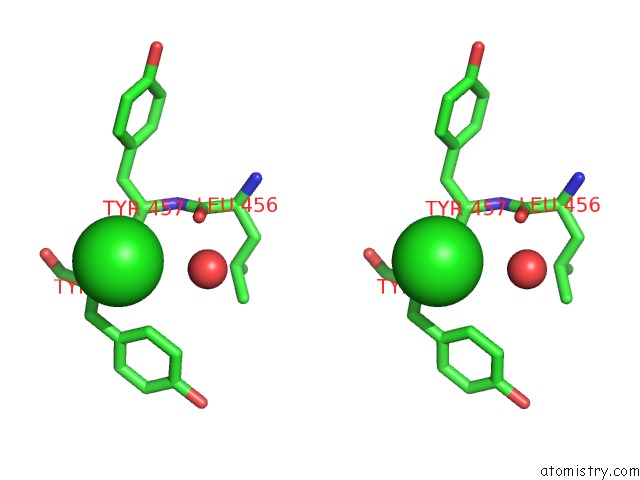
Stereo pair view
Mono view
Stereo pair view
A full contact list of Chlorine with other atoms in the Cl binding
site number 1 of Crystal Structure of Prefusion-Stabilized Rsv F Variant Sc-Tm within 5.0Å range:
|
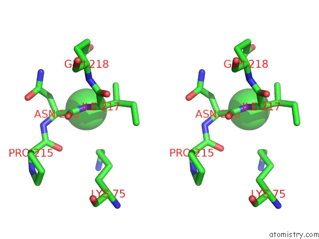
Chlorine binding site 2 out of 2 in 5c6b
Go back to
Chlorine binding site 2 out
of 2 in the Crystal Structure of Prefusion-Stabilized Rsv F Variant Sc-Tm
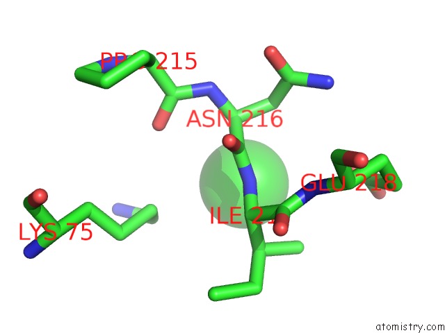
Mono view
Stereo pair view
Mono view
Stereo pair view
A full contact list of Chlorine with other atoms in the Cl binding
site number 2 of Crystal Structure of Prefusion-Stabilized Rsv F Variant Sc-Tm within 5.0Å range:
|
Reference:
A.Krarup,
D.Truan,
P.Furmanova-Hollenstein,
L.Bogaert,
P.Bouchier,
I.J.Bisschop,
M.N.Widjojoatmodjo,
R.Zahn,
H.Schuitemaker,
J.S.Mclellan,
J.P.Langedijk.
A Highly Stable Prefusion Rsv F Vaccine Derived From Structural Analysis of the Fusion Mechanism. Nat Commun V. 6 8143 2015.
ISSN: ESSN 2041-1723
PubMed: 26333350
DOI: 10.1038/NCOMMS9143
Page generated: Fri Jul 26 05:55:33 2024
ISSN: ESSN 2041-1723
PubMed: 26333350
DOI: 10.1038/NCOMMS9143
Last articles
Zn in 9J0NZn in 9J0O
Zn in 9J0P
Zn in 9FJX
Zn in 9EKB
Zn in 9C0F
Zn in 9CAH
Zn in 9CH0
Zn in 9CH3
Zn in 9CH1