Chlorine »
PDB 5cb7-5cni »
5cmk »
Chlorine in PDB 5cmk: Crystal Structure of the GLUK2EM Lbd Dimer Assembly Complex with Glutamate and LY466195
Protein crystallography data
The structure of Crystal Structure of the GLUK2EM Lbd Dimer Assembly Complex with Glutamate and LY466195, PDB code: 5cmk
was solved by
S.Chittori,
M.L.Mayer,
with X-Ray Crystallography technique. A brief refinement statistics is given in the table below:
| Resolution Low / High (Å) | 29.86 / 1.80 |
| Space group | P 61 2 2 |
| Cell size a, b, c (Å), α, β, γ (°) | 102.759, 102.759, 281.992, 90.00, 90.00, 120.00 |
| R / Rfree (%) | 16.4 / 18.7 |
Other elements in 5cmk:
The structure of Crystal Structure of the GLUK2EM Lbd Dimer Assembly Complex with Glutamate and LY466195 also contains other interesting chemical elements:
| Fluorine | (F) | 2 atoms |
Chlorine Binding Sites:
The binding sites of Chlorine atom in the Crystal Structure of the GLUK2EM Lbd Dimer Assembly Complex with Glutamate and LY466195
(pdb code 5cmk). This binding sites where shown within
5.0 Angstroms radius around Chlorine atom.
In total 2 binding sites of Chlorine where determined in the Crystal Structure of the GLUK2EM Lbd Dimer Assembly Complex with Glutamate and LY466195, PDB code: 5cmk:
Jump to Chlorine binding site number: 1; 2;
In total 2 binding sites of Chlorine where determined in the Crystal Structure of the GLUK2EM Lbd Dimer Assembly Complex with Glutamate and LY466195, PDB code: 5cmk:
Jump to Chlorine binding site number: 1; 2;
Chlorine binding site 1 out of 2 in 5cmk
Go back to
Chlorine binding site 1 out
of 2 in the Crystal Structure of the GLUK2EM Lbd Dimer Assembly Complex with Glutamate and LY466195
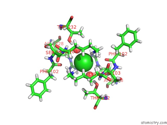
Mono view
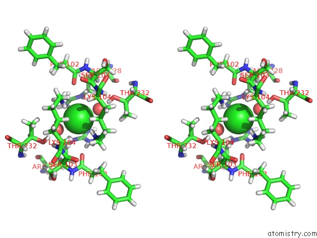
Stereo pair view
Mono view
Stereo pair view
A full contact list of Chlorine with other atoms in the Cl binding
site number 1 of Crystal Structure of the GLUK2EM Lbd Dimer Assembly Complex with Glutamate and LY466195 within 5.0Å range:
|
Chlorine binding site 2 out of 2 in 5cmk
Go back to
Chlorine binding site 2 out
of 2 in the Crystal Structure of the GLUK2EM Lbd Dimer Assembly Complex with Glutamate and LY466195
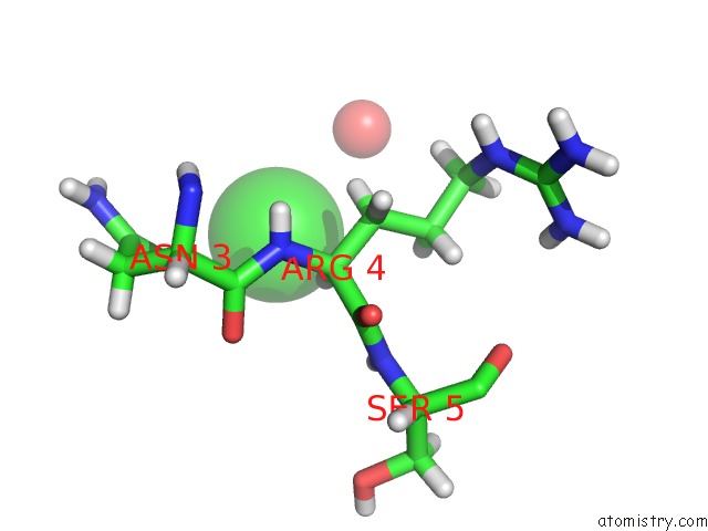
Mono view
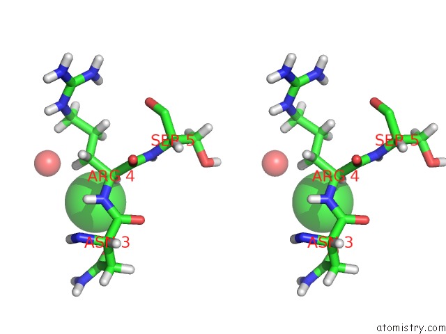
Stereo pair view
Mono view
Stereo pair view
A full contact list of Chlorine with other atoms in the Cl binding
site number 2 of Crystal Structure of the GLUK2EM Lbd Dimer Assembly Complex with Glutamate and LY466195 within 5.0Å range:
|
Reference:
J.R.Meyerson,
S.Chittori,
A.Merk,
P.Rao,
T.H.Han,
M.Serpe,
M.L.Mayer,
S.Subramaniam.
Structural Basis of Kainate Subtype Glutamate Receptor Desensitization. Nature V. 537 567 2016.
ISSN: ESSN 1476-4687
PubMed: 27580033
DOI: 10.1038/NATURE19352
Page generated: Sat Jul 12 00:50:09 2025
ISSN: ESSN 1476-4687
PubMed: 27580033
DOI: 10.1038/NATURE19352
Last articles
Fe in 2YXOFe in 2YRS
Fe in 2YXC
Fe in 2YNM
Fe in 2YVJ
Fe in 2YP1
Fe in 2YU2
Fe in 2YU1
Fe in 2YQB
Fe in 2YOO