Chlorine »
PDB 5cnm-5cu4 »
5cof »
Chlorine in PDB 5cof: Crystal Structure of Uncharacterised Protein Q1R1X2 From Escherichia Coli UTI89
Protein crystallography data
The structure of Crystal Structure of Uncharacterised Protein Q1R1X2 From Escherichia Coli UTI89, PDB code: 5cof
was solved by
J.D.Taylor,
S.Hare,
S.J.Matthews,
with X-Ray Crystallography technique. A brief refinement statistics is given in the table below:
| Resolution Low / High (Å) | 41.29 / 1.35 |
| Space group | P 21 21 21 |
| Cell size a, b, c (Å), α, β, γ (°) | 46.860, 48.160, 80.220, 90.00, 90.00, 90.00 |
| R / Rfree (%) | 11.5 / 13.8 |
Other elements in 5cof:
The structure of Crystal Structure of Uncharacterised Protein Q1R1X2 From Escherichia Coli UTI89 also contains other interesting chemical elements:
| Calcium | (Ca) | 2 atoms |
| Sodium | (Na) | 1 atom |
Chlorine Binding Sites:
The binding sites of Chlorine atom in the Crystal Structure of Uncharacterised Protein Q1R1X2 From Escherichia Coli UTI89
(pdb code 5cof). This binding sites where shown within
5.0 Angstroms radius around Chlorine atom.
In total only one binding site of Chlorine was determined in the Crystal Structure of Uncharacterised Protein Q1R1X2 From Escherichia Coli UTI89, PDB code: 5cof:
In total only one binding site of Chlorine was determined in the Crystal Structure of Uncharacterised Protein Q1R1X2 From Escherichia Coli UTI89, PDB code: 5cof:
Chlorine binding site 1 out of 1 in 5cof
Go back to
Chlorine binding site 1 out
of 1 in the Crystal Structure of Uncharacterised Protein Q1R1X2 From Escherichia Coli UTI89

Mono view
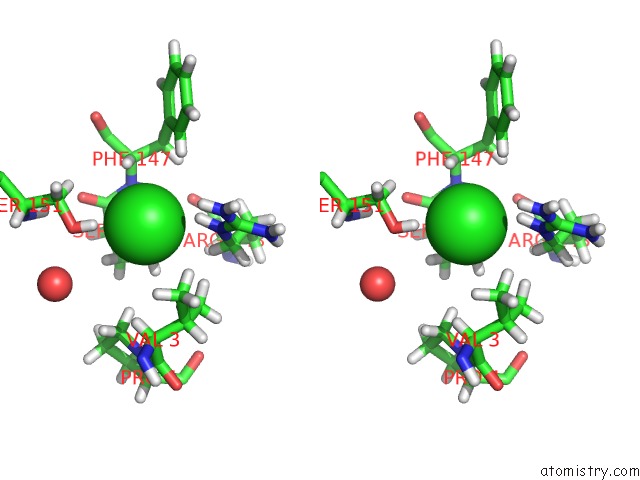
Stereo pair view
Mono view
Stereo pair view
A full contact list of Chlorine with other atoms in the Cl binding
site number 1 of Crystal Structure of Uncharacterised Protein Q1R1X2 From Escherichia Coli UTI89 within 5.0Å range:
|
Reference:
J.D.Taylor,
G.Taylor,
S.A.Hare,
S.J.Matthews.
Structures of the Dfsb Protein Family Suggest A Cationic, Helical Sibling Lethal Factor Peptide. J.Mol.Biol. V. 428 554 2016.
ISSN: ESSN 1089-8638
PubMed: 26804569
DOI: 10.1016/J.JMB.2016.01.013
Page generated: Sat Jul 12 00:53:09 2025
ISSN: ESSN 1089-8638
PubMed: 26804569
DOI: 10.1016/J.JMB.2016.01.013
Last articles
Fe in 2YXOFe in 2YRS
Fe in 2YXC
Fe in 2YNM
Fe in 2YVJ
Fe in 2YP1
Fe in 2YU2
Fe in 2YU1
Fe in 2YQB
Fe in 2YOO