Chlorine »
PDB 5cnm-5cu4 »
5ctu »
Chlorine in PDB 5ctu: Crystal Structure of the Atp Binding Domain of S. Aureus Gyrb Complexed with A Fragment
Enzymatic activity of Crystal Structure of the Atp Binding Domain of S. Aureus Gyrb Complexed with A Fragment
All present enzymatic activity of Crystal Structure of the Atp Binding Domain of S. Aureus Gyrb Complexed with A Fragment:
5.99.1.3;
5.99.1.3;
Protein crystallography data
The structure of Crystal Structure of the Atp Binding Domain of S. Aureus Gyrb Complexed with A Fragment, PDB code: 5ctu
was solved by
O.A.Andersen,
J.Barker,
R.K.Cheng,
J.Kahmann,
B.Felicetti,
M.Wood,
C.Scheich,
M.Mesleh,
J.B.Cross,
J.Zhang,
Q.Yang,
B.Lippa,
M.D.Ryan,
with X-Ray Crystallography technique. A brief refinement statistics is given in the table below:
| Resolution Low / High (Å) | 25.86 / 1.45 |
| Space group | C 1 2 1 |
| Cell size a, b, c (Å), α, β, γ (°) | 142.660, 55.650, 51.140, 90.00, 101.01, 90.00 |
| R / Rfree (%) | 17.6 / 20.4 |
Other elements in 5ctu:
The structure of Crystal Structure of the Atp Binding Domain of S. Aureus Gyrb Complexed with A Fragment also contains other interesting chemical elements:
| Magnesium | (Mg) | 1 atom |
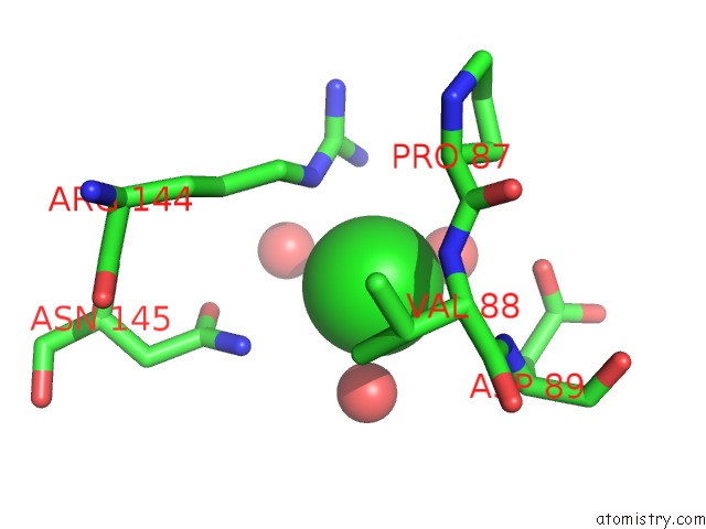
Chlorine Binding Sites:
The binding sites of Chlorine atom in the Crystal Structure of the Atp Binding Domain of S. Aureus Gyrb Complexed with A Fragment
(pdb code 5ctu). This binding sites where shown within
5.0 Angstroms radius around Chlorine atom.
In total only one binding site of Chlorine was determined in the Crystal Structure of the Atp Binding Domain of S. Aureus Gyrb Complexed with A Fragment, PDB code: 5ctu:
In total only one binding site of Chlorine was determined in the Crystal Structure of the Atp Binding Domain of S. Aureus Gyrb Complexed with A Fragment, PDB code: 5ctu:
Chlorine binding site 1 out of 1 in 5ctu
Go back to
Chlorine binding site 1 out
of 1 in the Crystal Structure of the Atp Binding Domain of S. Aureus Gyrb Complexed with A Fragment
Mono view

Stereo pair view
Mono view
Stereo pair view
A full contact list of Chlorine with other atoms in the Cl binding
site number 1 of Crystal Structure of the Atp Binding Domain of S. Aureus Gyrb Complexed with A Fragment within 5.0Å range:
|
Reference:
M.F.Mesleh,
J.B.Cross,
J.Zhang,
J.Kahmann,
O.A.Andersen,
J.Barker,
R.K.Cheng,
B.Felicetti,
M.Wood,
A.T.Hadfield,
C.Scheich,
T.I.Moy,
Q.Yang,
J.Shotwell,
K.Nguyen,
B.Lippa,
R.Dolle,
M.D.Ryan.
Fragment-Based Discovery of Dna Gyrase Inhibitors Targeting the Atpase Subunit of Gyrb. Bioorg.Med.Chem.Lett. V. 26 1314 2016.
ISSN: ESSN 1464-3405
PubMed: 26786695
DOI: 10.1016/J.BMCL.2016.01.009
Page generated: Fri Jul 26 06:16:38 2024
ISSN: ESSN 1464-3405
PubMed: 26786695
DOI: 10.1016/J.BMCL.2016.01.009
Last articles
Zn in 9J0NZn in 9J0O
Zn in 9J0P
Zn in 9FJX
Zn in 9EKB
Zn in 9C0F
Zn in 9CAH
Zn in 9CH0
Zn in 9CH3
Zn in 9CH1