Chlorine »
PDB 5cu8-5d0x »
5cyv »
Chlorine in PDB 5cyv: Crystal Structure of Cour From Rhodococcus Jostii RHA1 Bound to P- Coumaroyl-Coa
Protein crystallography data
The structure of Crystal Structure of Cour From Rhodococcus Jostii RHA1 Bound to P- Coumaroyl-Coa, PDB code: 5cyv
was solved by
P.J.Stogios,
X.Xu,
A.Dong,
A.Savchenko,
A.Joachimiak,
Midwest Center Forstructural Genomics (Mcsg),
with X-Ray Crystallography technique. A brief refinement statistics is given in the table below:
| Resolution Low / High (Å) | 24.79 / 1.52 |
| Space group | C 2 2 21 |
| Cell size a, b, c (Å), α, β, γ (°) | 64.145, 134.385, 73.336, 90.00, 90.00, 90.00 |
| R / Rfree (%) | 16.1 / 20.2 |
Other elements in 5cyv:
The structure of Crystal Structure of Cour From Rhodococcus Jostii RHA1 Bound to P- Coumaroyl-Coa also contains other interesting chemical elements:
| Magnesium | (Mg) | 3 atoms |
Chlorine Binding Sites:
The binding sites of Chlorine atom in the Crystal Structure of Cour From Rhodococcus Jostii RHA1 Bound to P- Coumaroyl-Coa
(pdb code 5cyv). This binding sites where shown within
5.0 Angstroms radius around Chlorine atom.
In total 3 binding sites of Chlorine where determined in the Crystal Structure of Cour From Rhodococcus Jostii RHA1 Bound to P- Coumaroyl-Coa, PDB code: 5cyv:
Jump to Chlorine binding site number: 1; 2; 3;
In total 3 binding sites of Chlorine where determined in the Crystal Structure of Cour From Rhodococcus Jostii RHA1 Bound to P- Coumaroyl-Coa, PDB code: 5cyv:
Jump to Chlorine binding site number: 1; 2; 3;
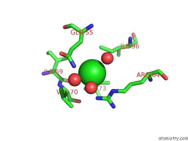
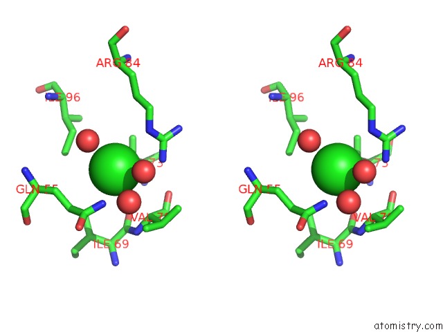
Chlorine binding site 1 out of 3 in 5cyv
Go back to
Chlorine binding site 1 out
of 3 in the Crystal Structure of Cour From Rhodococcus Jostii RHA1 Bound to P- Coumaroyl-Coa
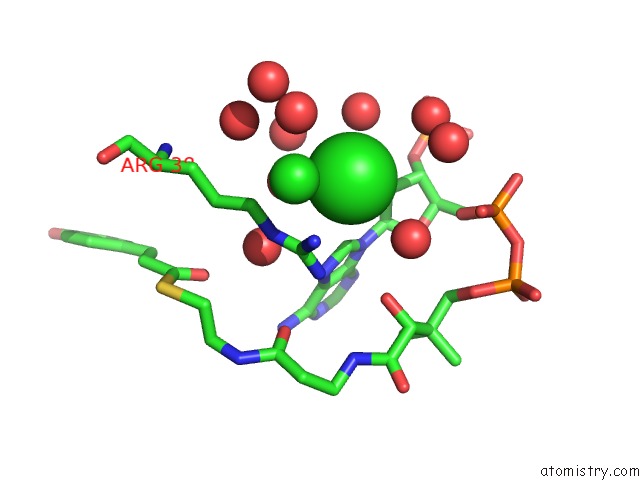
Mono view
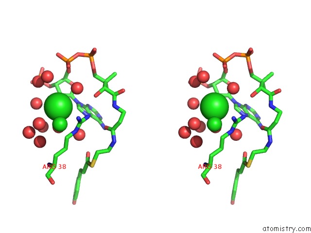
Stereo pair view
Mono view
Stereo pair view
A full contact list of Chlorine with other atoms in the Cl binding
site number 1 of Crystal Structure of Cour From Rhodococcus Jostii RHA1 Bound to P- Coumaroyl-Coa within 5.0Å range:
|
Chlorine binding site 2 out of 3 in 5cyv
Go back to
Chlorine binding site 2 out
of 3 in the Crystal Structure of Cour From Rhodococcus Jostii RHA1 Bound to P- Coumaroyl-Coa
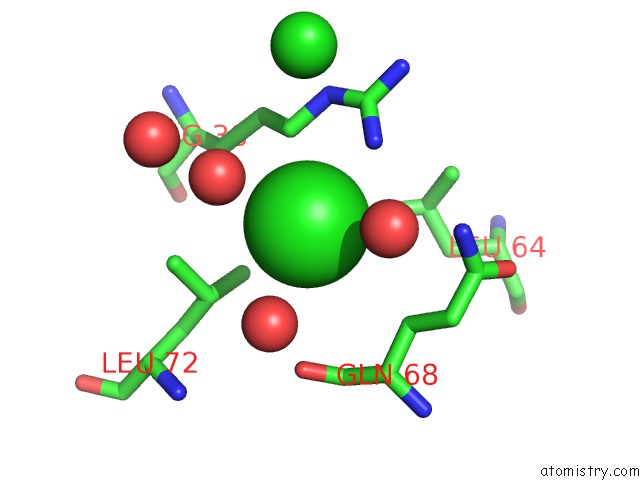
Mono view
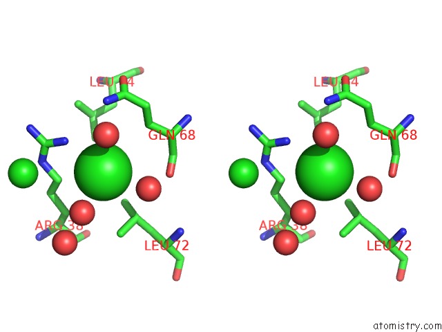
Stereo pair view
Mono view
Stereo pair view
A full contact list of Chlorine with other atoms in the Cl binding
site number 2 of Crystal Structure of Cour From Rhodococcus Jostii RHA1 Bound to P- Coumaroyl-Coa within 5.0Å range:
|
Chlorine binding site 3 out of 3 in 5cyv
Go back to
Chlorine binding site 3 out
of 3 in the Crystal Structure of Cour From Rhodococcus Jostii RHA1 Bound to P- Coumaroyl-Coa
Mono view
Stereo pair view
Mono view
Stereo pair view
A full contact list of Chlorine with other atoms in the Cl binding
site number 3 of Crystal Structure of Cour From Rhodococcus Jostii RHA1 Bound to P- Coumaroyl-Coa within 5.0Å range:
|
Reference:
H.Otani,
P.J.Stogios,
X.Xu,
B.Nocek,
S.N.Li,
A.Savchenko,
L.D.Eltis.
The Activity of Cour, A Marr Family Transcriptional Regulator, Is Modulated Through A Novel Molecular Mechanism. Nucleic Acids Res. V. 44 595 2016.
ISSN: ESSN 1362-4962
PubMed: 26400178
DOI: 10.1093/NAR/GKV955
Page generated: Fri Jul 26 06:21:14 2024
ISSN: ESSN 1362-4962
PubMed: 26400178
DOI: 10.1093/NAR/GKV955
Last articles
Zn in 9J0NZn in 9J0O
Zn in 9J0P
Zn in 9FJX
Zn in 9EKB
Zn in 9C0F
Zn in 9CAH
Zn in 9CH0
Zn in 9CH3
Zn in 9CH1