Chlorine »
PDB 5dab-5dij »
5df7 »
Chlorine in PDB 5df7: Crystal Structure of Penicillin-Binding Protein 3 From Pseudomonas Aeruginosa in Complex with Azlocillin
Enzymatic activity of Crystal Structure of Penicillin-Binding Protein 3 From Pseudomonas Aeruginosa in Complex with Azlocillin
All present enzymatic activity of Crystal Structure of Penicillin-Binding Protein 3 From Pseudomonas Aeruginosa in Complex with Azlocillin:
2.4.1.129;
2.4.1.129;
Protein crystallography data
The structure of Crystal Structure of Penicillin-Binding Protein 3 From Pseudomonas Aeruginosa in Complex with Azlocillin, PDB code: 5df7
was solved by
J.Ren,
J.E.Nettleship,
A.Males,
D.I.Stuart,
R.J.Owens,
with X-Ray Crystallography technique. A brief refinement statistics is given in the table below:
| Resolution Low / High (Å) | 47.19 / 2.00 |
| Space group | P 1 |
| Cell size a, b, c (Å), α, β, γ (°) | 57.253, 74.919, 82.722, 71.26, 85.99, 85.69 |
| R / Rfree (%) | 19.8 / 23.7 |
Chlorine Binding Sites:
The binding sites of Chlorine atom in the Crystal Structure of Penicillin-Binding Protein 3 From Pseudomonas Aeruginosa in Complex with Azlocillin
(pdb code 5df7). This binding sites where shown within
5.0 Angstroms radius around Chlorine atom.
In total 4 binding sites of Chlorine where determined in the Crystal Structure of Penicillin-Binding Protein 3 From Pseudomonas Aeruginosa in Complex with Azlocillin, PDB code: 5df7:
Jump to Chlorine binding site number: 1; 2; 3; 4;
In total 4 binding sites of Chlorine where determined in the Crystal Structure of Penicillin-Binding Protein 3 From Pseudomonas Aeruginosa in Complex with Azlocillin, PDB code: 5df7:
Jump to Chlorine binding site number: 1; 2; 3; 4;
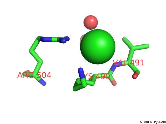
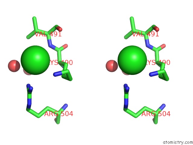
Chlorine binding site 1 out of 4 in 5df7
Go back to
Chlorine binding site 1 out
of 4 in the Crystal Structure of Penicillin-Binding Protein 3 From Pseudomonas Aeruginosa in Complex with Azlocillin
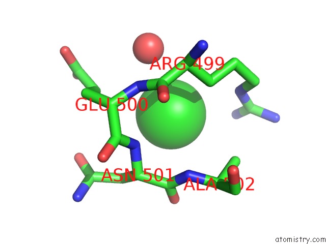
Mono view
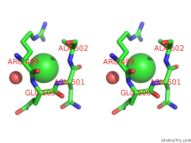
Stereo pair view
Mono view
Stereo pair view
A full contact list of Chlorine with other atoms in the Cl binding
site number 1 of Crystal Structure of Penicillin-Binding Protein 3 From Pseudomonas Aeruginosa in Complex with Azlocillin within 5.0Å range:
|
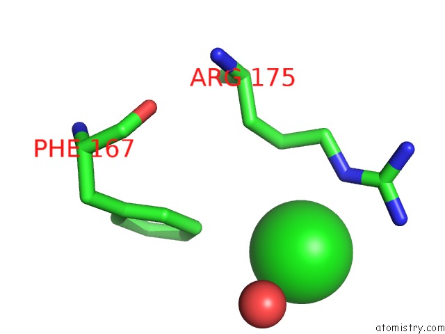
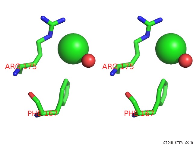
Chlorine binding site 2 out of 4 in 5df7
Go back to
Chlorine binding site 2 out
of 4 in the Crystal Structure of Penicillin-Binding Protein 3 From Pseudomonas Aeruginosa in Complex with Azlocillin
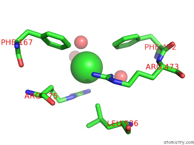
Mono view
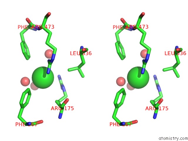
Stereo pair view
Mono view
Stereo pair view
A full contact list of Chlorine with other atoms in the Cl binding
site number 2 of Crystal Structure of Penicillin-Binding Protein 3 From Pseudomonas Aeruginosa in Complex with Azlocillin within 5.0Å range:
|
Chlorine binding site 3 out of 4 in 5df7
Go back to
Chlorine binding site 3 out
of 4 in the Crystal Structure of Penicillin-Binding Protein 3 From Pseudomonas Aeruginosa in Complex with Azlocillin
Mono view
Stereo pair view
Mono view
Stereo pair view
A full contact list of Chlorine with other atoms in the Cl binding
site number 3 of Crystal Structure of Penicillin-Binding Protein 3 From Pseudomonas Aeruginosa in Complex with Azlocillin within 5.0Å range:
|
Chlorine binding site 4 out of 4 in 5df7
Go back to
Chlorine binding site 4 out
of 4 in the Crystal Structure of Penicillin-Binding Protein 3 From Pseudomonas Aeruginosa in Complex with Azlocillin
Mono view
Stereo pair view
Mono view
Stereo pair view
A full contact list of Chlorine with other atoms in the Cl binding
site number 4 of Crystal Structure of Penicillin-Binding Protein 3 From Pseudomonas Aeruginosa in Complex with Azlocillin within 5.0Å range:
|
Reference:
J.Ren,
J.E.Nettleship,
A.Males,
D.I.Stuart,
R.J.Owens.
Crystal Structures of Penicillin-Binding Protein 3 in Complexes with Azlocillin and Cefoperazone in Both Acylated and Deacylated Forms. Febs Lett. V. 590 288 2016.
ISSN: ISSN 0014-5793
PubMed: 26823174
DOI: 10.1002/1873-3468.12054
Page generated: Fri Jul 26 06:39:58 2024
ISSN: ISSN 0014-5793
PubMed: 26823174
DOI: 10.1002/1873-3468.12054
Last articles
Zn in 9J0NZn in 9J0O
Zn in 9J0P
Zn in 9FJX
Zn in 9EKB
Zn in 9C0F
Zn in 9CAH
Zn in 9CH0
Zn in 9CH3
Zn in 9CH1