Chlorine »
PDB 5dj1-5dtj »
5dn9 »
Chlorine in PDB 5dn9: Crystal Structure of Candida Boidinii Formate Dehydrogenase Complexed with Nad+ and Azide
Enzymatic activity of Crystal Structure of Candida Boidinii Formate Dehydrogenase Complexed with Nad+ and Azide
All present enzymatic activity of Crystal Structure of Candida Boidinii Formate Dehydrogenase Complexed with Nad+ and Azide:
1.2.1.2;
1.2.1.2;
Protein crystallography data
The structure of Crystal Structure of Candida Boidinii Formate Dehydrogenase Complexed with Nad+ and Azide, PDB code: 5dn9
was solved by
Q.Guo,
L.Gakhar,
K.Wichersham,
K.Francis,
A.Vardi-Kilshtain,
D.T.Major,
C.M.Cheatum,
A.Kohen,
with X-Ray Crystallography technique. A brief refinement statistics is given in the table below:
| Resolution Low / High (Å) | 41.98 / 1.50 |
| Space group | P 1 21 1 |
| Cell size a, b, c (Å), α, β, γ (°) | 50.931, 116.617, 63.208, 90.00, 106.90, 90.00 |
| R / Rfree (%) | 13.8 / 16.9 |
Chlorine Binding Sites:
The binding sites of Chlorine atom in the Crystal Structure of Candida Boidinii Formate Dehydrogenase Complexed with Nad+ and Azide
(pdb code 5dn9). This binding sites where shown within
5.0 Angstroms radius around Chlorine atom.
In total 2 binding sites of Chlorine where determined in the Crystal Structure of Candida Boidinii Formate Dehydrogenase Complexed with Nad+ and Azide, PDB code: 5dn9:
Jump to Chlorine binding site number: 1; 2;
In total 2 binding sites of Chlorine where determined in the Crystal Structure of Candida Boidinii Formate Dehydrogenase Complexed with Nad+ and Azide, PDB code: 5dn9:
Jump to Chlorine binding site number: 1; 2;
Chlorine binding site 1 out of 2 in 5dn9
Go back to
Chlorine binding site 1 out
of 2 in the Crystal Structure of Candida Boidinii Formate Dehydrogenase Complexed with Nad+ and Azide
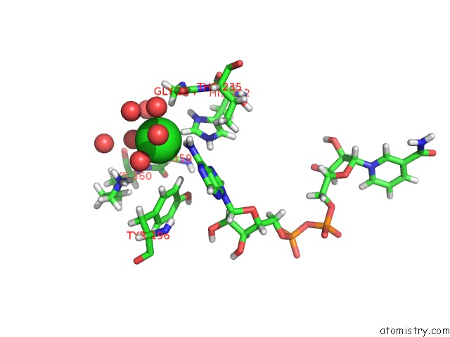
Mono view
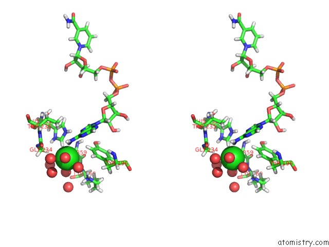
Stereo pair view
Mono view
Stereo pair view
A full contact list of Chlorine with other atoms in the Cl binding
site number 1 of Crystal Structure of Candida Boidinii Formate Dehydrogenase Complexed with Nad+ and Azide within 5.0Å range:
|
Chlorine binding site 2 out of 2 in 5dn9
Go back to
Chlorine binding site 2 out
of 2 in the Crystal Structure of Candida Boidinii Formate Dehydrogenase Complexed with Nad+ and Azide
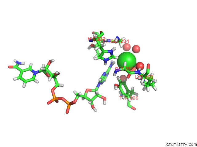
Mono view
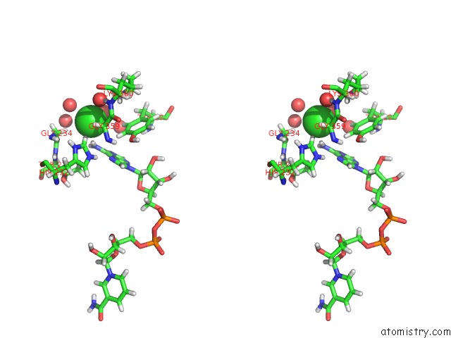
Stereo pair view
Mono view
Stereo pair view
A full contact list of Chlorine with other atoms in the Cl binding
site number 2 of Crystal Structure of Candida Boidinii Formate Dehydrogenase Complexed with Nad+ and Azide within 5.0Å range:
|
Reference:
Q.Guo,
L.Gakhar,
K.Wickersham,
K.Francis,
A.Vardi-Kilshtain,
D.T.Major,
C.M.Cheatum,
A.Kohen.
Structural and Kinetic Studies of Formate Dehydrogenase From Candida Boidinii. Biochemistry V. 55 2760 2016.
ISSN: ISSN 0006-2960
PubMed: 27100912
DOI: 10.1021/ACS.BIOCHEM.6B00181
Page generated: Sat Jul 12 01:19:29 2025
ISSN: ISSN 0006-2960
PubMed: 27100912
DOI: 10.1021/ACS.BIOCHEM.6B00181
Last articles
Fe in 2YXOFe in 2YRS
Fe in 2YXC
Fe in 2YNM
Fe in 2YVJ
Fe in 2YP1
Fe in 2YU2
Fe in 2YU1
Fe in 2YQB
Fe in 2YOO