Chlorine »
PDB 5e20-5eae »
5e75 »
Chlorine in PDB 5e75: Crystal Structure of BACOVA_02651
Protein crystallography data
The structure of Crystal Structure of BACOVA_02651, PDB code: 5e75
was solved by
N.M.Koropatkin,
with X-Ray Crystallography technique. A brief refinement statistics is given in the table below:
| Resolution Low / High (Å) | 21.48 / 1.36 |
| Space group | P 1 21 1 |
| Cell size a, b, c (Å), α, β, γ (°) | 52.800, 81.410, 57.680, 90.00, 107.85, 90.00 |
| R / Rfree (%) | 14.7 / 17.4 |
Other elements in 5e75:
The structure of Crystal Structure of BACOVA_02651 also contains other interesting chemical elements:
| Magnesium | (Mg) | 2 atoms |
Chlorine Binding Sites:
The binding sites of Chlorine atom in the Crystal Structure of BACOVA_02651
(pdb code 5e75). This binding sites where shown within
5.0 Angstroms radius around Chlorine atom.
In total 2 binding sites of Chlorine where determined in the Crystal Structure of BACOVA_02651, PDB code: 5e75:
Jump to Chlorine binding site number: 1; 2;
In total 2 binding sites of Chlorine where determined in the Crystal Structure of BACOVA_02651, PDB code: 5e75:
Jump to Chlorine binding site number: 1; 2;
Chlorine binding site 1 out of 2 in 5e75
Go back to
Chlorine binding site 1 out
of 2 in the Crystal Structure of BACOVA_02651
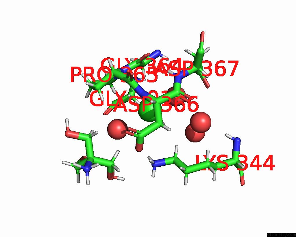
Mono view
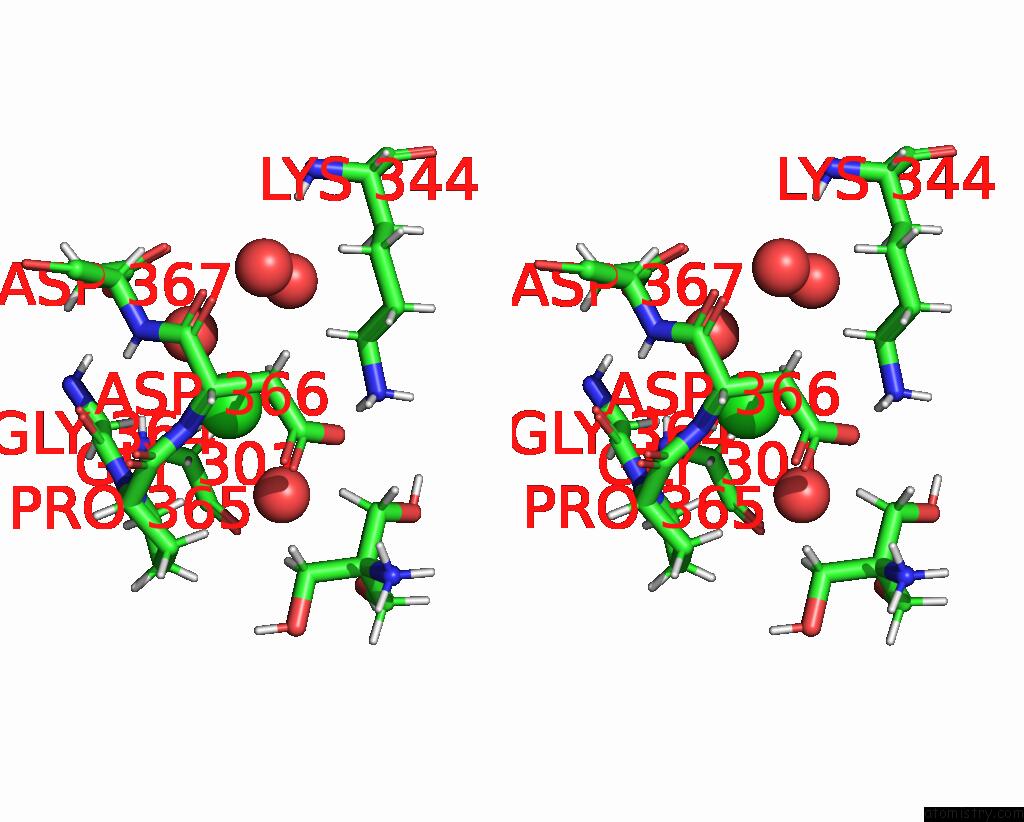
Stereo pair view
Mono view
Stereo pair view
A full contact list of Chlorine with other atoms in the Cl binding
site number 1 of Crystal Structure of BACOVA_02651 within 5.0Å range:
|
Chlorine binding site 2 out of 2 in 5e75
Go back to
Chlorine binding site 2 out
of 2 in the Crystal Structure of BACOVA_02651
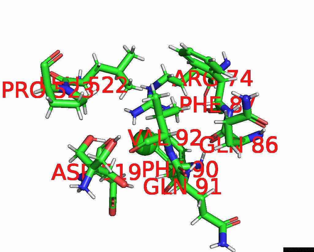
Mono view

Stereo pair view
Mono view
Stereo pair view
A full contact list of Chlorine with other atoms in the Cl binding
site number 2 of Crystal Structure of BACOVA_02651 within 5.0Å range:
|
Reference:
A.S.Tauzin,
K.J.Kwiatkowski,
N.I.Orlovsky,
C.J.Smith,
A.L.Creagh,
C.A.Haynes,
Z.Wawrzak,
H.Brumer,
N.M.Koropatkin.
Molecular Dissection of Xyloglucan Recognition in A Prominent Human Gut Symbiont. Mbio V. 7 02134 2016.
ISSN: ESSN 2150-7511
PubMed: 27118585
DOI: 10.1128/MBIO.02134-15
Page generated: Fri Jul 26 07:06:33 2024
ISSN: ESSN 2150-7511
PubMed: 27118585
DOI: 10.1128/MBIO.02134-15
Last articles
Zn in 9J0NZn in 9J0O
Zn in 9J0P
Zn in 9FJX
Zn in 9EKB
Zn in 9C0F
Zn in 9CAH
Zn in 9CH0
Zn in 9CH3
Zn in 9CH1