Chlorine »
PDB 5e20-5eae »
5e89 »
Chlorine in PDB 5e89: Crystal Structure of Human Galectin-3 Crd in Complex with 3- Fluophenyl-1,2,3-Triazolyl Thiodigalactoside Inhibitor
Protein crystallography data
The structure of Crystal Structure of Human Galectin-3 Crd in Complex with 3- Fluophenyl-1,2,3-Triazolyl Thiodigalactoside Inhibitor, PDB code: 5e89
was solved by
P.M.Collins,
H.Blanchard,
with X-Ray Crystallography technique. A brief refinement statistics is given in the table below:
| Resolution Low / High (Å) | 43.01 / 1.50 |
| Space group | P 21 21 21 |
| Cell size a, b, c (Å), α, β, γ (°) | 37.045, 58.298, 63.728, 90.00, 90.00, 90.00 |
| R / Rfree (%) | 16.8 / 19 |
Other elements in 5e89:
The structure of Crystal Structure of Human Galectin-3 Crd in Complex with 3- Fluophenyl-1,2,3-Triazolyl Thiodigalactoside Inhibitor also contains other interesting chemical elements:
| Fluorine | (F) | 2 atoms |
Chlorine Binding Sites:
The binding sites of Chlorine atom in the Crystal Structure of Human Galectin-3 Crd in Complex with 3- Fluophenyl-1,2,3-Triazolyl Thiodigalactoside Inhibitor
(pdb code 5e89). This binding sites where shown within
5.0 Angstroms radius around Chlorine atom.
In total only one binding site of Chlorine was determined in the Crystal Structure of Human Galectin-3 Crd in Complex with 3- Fluophenyl-1,2,3-Triazolyl Thiodigalactoside Inhibitor, PDB code: 5e89:
In total only one binding site of Chlorine was determined in the Crystal Structure of Human Galectin-3 Crd in Complex with 3- Fluophenyl-1,2,3-Triazolyl Thiodigalactoside Inhibitor, PDB code: 5e89:
Chlorine binding site 1 out of 1 in 5e89
Go back to
Chlorine binding site 1 out
of 1 in the Crystal Structure of Human Galectin-3 Crd in Complex with 3- Fluophenyl-1,2,3-Triazolyl Thiodigalactoside Inhibitor
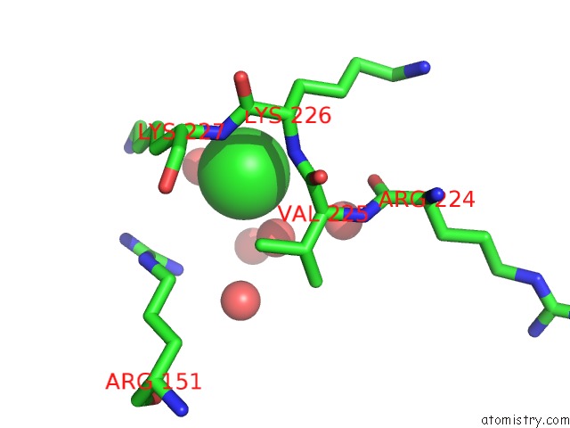
Mono view
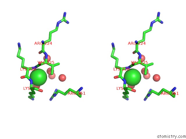
Stereo pair view
Mono view
Stereo pair view
A full contact list of Chlorine with other atoms in the Cl binding
site number 1 of Crystal Structure of Human Galectin-3 Crd in Complex with 3- Fluophenyl-1,2,3-Triazolyl Thiodigalactoside Inhibitor within 5.0Å range:
|
Reference:
T.Delaine,
P.Collins,
A.Mackinnon,
G.Sharma,
J.Stegmayr,
V.K.Rajput,
S.Mandal,
I.Cumpstey,
A.Larumbe,
B.A.Salameh,
B.Kahl-Knutsson,
H.Van Hattum,
M.Van Scherpenzeel,
R.J.Pieters,
T.Sethi,
H.Schambye,
S.Oredsson,
H.Leffler,
H.Blanchard,
U.J.Nilsson.
Galectin-3-Binding Glycomimetics That Strongly Reduce Bleomycin-Induced Lung Fibrosis and Modulate Intracellular Glycan Recognition. Chembiochem V. 17 1759 2016.
ISSN: ESSN 1439-7633
PubMed: 27356186
DOI: 10.1002/CBIC.201600285
Page generated: Fri Jul 26 07:08:16 2024
ISSN: ESSN 1439-7633
PubMed: 27356186
DOI: 10.1002/CBIC.201600285
Last articles
Zn in 9J0NZn in 9J0O
Zn in 9J0P
Zn in 9FJX
Zn in 9EKB
Zn in 9C0F
Zn in 9CAH
Zn in 9CH0
Zn in 9CH3
Zn in 9CH1