Chlorine »
PDB 5e20-5eae »
5e9c »
Chlorine in PDB 5e9c: Crystal Structure of Human Heparanase in Complex with Heparin Tetrasaccharide DP4
Enzymatic activity of Crystal Structure of Human Heparanase in Complex with Heparin Tetrasaccharide DP4
All present enzymatic activity of Crystal Structure of Human Heparanase in Complex with Heparin Tetrasaccharide DP4:
3.2.1.166;
3.2.1.166;
Protein crystallography data
The structure of Crystal Structure of Human Heparanase in Complex with Heparin Tetrasaccharide DP4, PDB code: 5e9c
was solved by
L.Wu,
G.J.Davies,
with X-Ray Crystallography technique. A brief refinement statistics is given in the table below:
| Resolution Low / High (Å) | 78.27 / 1.73 |
| Space group | P 1 21 1 |
| Cell size a, b, c (Å), α, β, γ (°) | 46.220, 71.120, 78.600, 90.00, 95.23, 90.00 |
| R / Rfree (%) | 17.2 / 21.1 |
Chlorine Binding Sites:
The binding sites of Chlorine atom in the Crystal Structure of Human Heparanase in Complex with Heparin Tetrasaccharide DP4
(pdb code 5e9c). This binding sites where shown within
5.0 Angstroms radius around Chlorine atom.
In total only one binding site of Chlorine was determined in the Crystal Structure of Human Heparanase in Complex with Heparin Tetrasaccharide DP4, PDB code: 5e9c:
In total only one binding site of Chlorine was determined in the Crystal Structure of Human Heparanase in Complex with Heparin Tetrasaccharide DP4, PDB code: 5e9c:
Chlorine binding site 1 out of 1 in 5e9c
Go back to
Chlorine binding site 1 out
of 1 in the Crystal Structure of Human Heparanase in Complex with Heparin Tetrasaccharide DP4
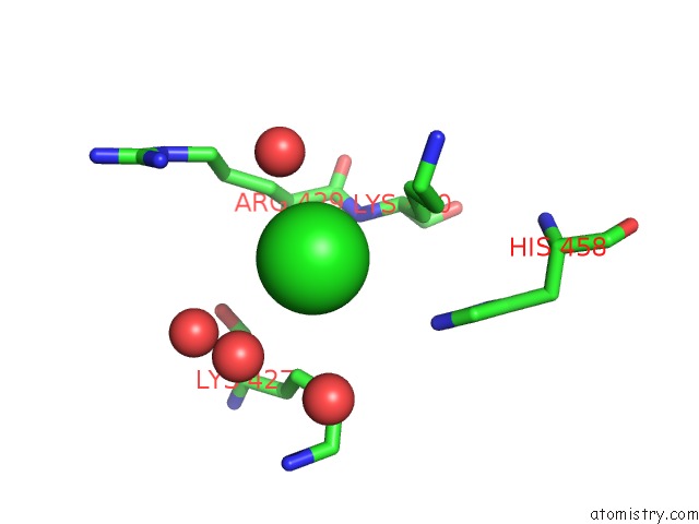
Mono view
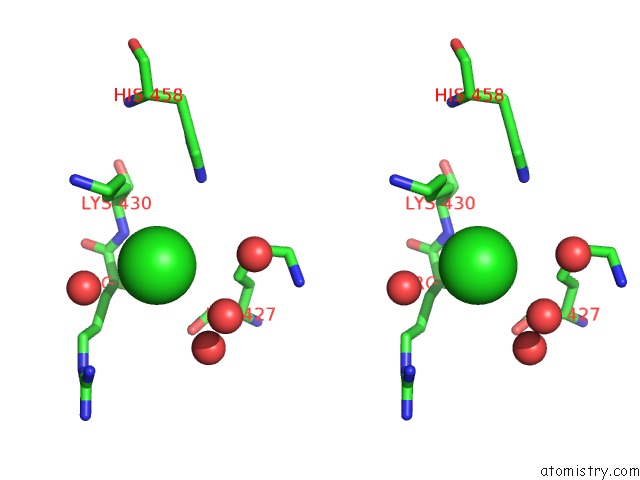
Stereo pair view
Mono view
Stereo pair view
A full contact list of Chlorine with other atoms in the Cl binding
site number 1 of Crystal Structure of Human Heparanase in Complex with Heparin Tetrasaccharide DP4 within 5.0Å range:
|
Reference:
L.Wu,
C.M.Viola,
A.M.Brzozowski,
G.J.Davies.
Structural Characterization of Human Heparanase Reveals Insights Into Substrate Recognition. Nat.Struct.Mol.Biol. V. 22 1016 2015.
ISSN: ESSN 1545-9985
PubMed: 26575439
DOI: 10.1038/NSMB.3136
Page generated: Sat Jul 12 01:35:33 2025
ISSN: ESSN 1545-9985
PubMed: 26575439
DOI: 10.1038/NSMB.3136
Last articles
Fe in 2YXOFe in 2YRS
Fe in 2YXC
Fe in 2YNM
Fe in 2YVJ
Fe in 2YP1
Fe in 2YU2
Fe in 2YU1
Fe in 2YQB
Fe in 2YOO