Chlorine in PDB 5eoe: Crystal Structure of Extended-Spectrum Beta-Lactamase Bel-1 (Orthorhombic Form)
Enzymatic activity of Crystal Structure of Extended-Spectrum Beta-Lactamase Bel-1 (Orthorhombic Form)
All present enzymatic activity of Crystal Structure of Extended-Spectrum Beta-Lactamase Bel-1 (Orthorhombic Form):
3.5.2.6;
Protein crystallography data
The structure of Crystal Structure of Extended-Spectrum Beta-Lactamase Bel-1 (Orthorhombic Form), PDB code: 5eoe
was solved by
C.Pozzi,
F.De Luca,
M.Benvenuti,
J.D.Docquier,
S.Mangani,
with X-Ray Crystallography technique. A brief refinement statistics is given in the table below:
Resolution Low / High (Å)
|
29.25 /
1.60
|
Space group
|
P 21 21 2
|
Cell size a, b, c (Å), α, β, γ (°)
|
100.270,
120.920,
53.410,
90.00,
90.00,
90.00
|
R / Rfree (%)
|
14.2 /
16.7
|
Chlorine Binding Sites:
The binding sites of Chlorine atom in the Crystal Structure of Extended-Spectrum Beta-Lactamase Bel-1 (Orthorhombic Form)
(pdb code 5eoe). This binding sites where shown within
5.0 Angstroms radius around Chlorine atom.
In total 6 binding sites of Chlorine where determined in the
Crystal Structure of Extended-Spectrum Beta-Lactamase Bel-1 (Orthorhombic Form), PDB code: 5eoe:
Jump to Chlorine binding site number:
1;
2;
3;
4;
5;
6;
Chlorine binding site 1 out
of 6 in 5eoe
Go back to
Chlorine Binding Sites List in 5eoe
Chlorine binding site 1 out
of 6 in the Crystal Structure of Extended-Spectrum Beta-Lactamase Bel-1 (Orthorhombic Form)
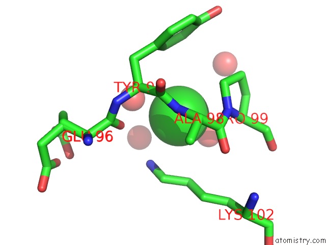
Mono view
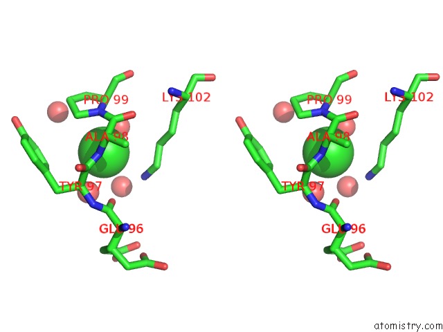
Stereo pair view
|
A full contact list of Chlorine with other atoms in the Cl binding
site number 1 of Crystal Structure of Extended-Spectrum Beta-Lactamase Bel-1 (Orthorhombic Form) within 5.0Å range:
probe
|
atom
|
residue
|
distance (Å)
|
B
|
Occ
|
A:Cl301
b:18.8
occ:1.00
|
O
|
B:HOH759
|
2.9
|
23.3
|
1.0
|
O
|
A:HOH568
|
3.1
|
38.7
|
1.0
|
N
|
A:ALA98
|
3.2
|
9.7
|
1.0
|
NZ
|
A:LYS102
|
3.2
|
14.2
|
1.0
|
N
|
A:PRO99
|
3.5
|
9.7
|
1.0
|
O
|
A:GLU96
|
3.6
|
13.9
|
1.0
|
CA
|
A:PRO99
|
3.7
|
9.6
|
1.0
|
CD
|
A:PRO99
|
3.8
|
10.1
|
1.0
|
C
|
A:ALA98
|
3.8
|
9.5
|
1.0
|
CA
|
A:TYR97
|
3.8
|
10.6
|
1.0
|
CE
|
A:LYS102
|
3.8
|
13.2
|
1.0
|
CD
|
A:LYS102
|
3.9
|
12.2
|
1.0
|
O
|
A:HOH672
|
3.9
|
46.3
|
1.0
|
C
|
A:TYR97
|
4.0
|
9.9
|
1.0
|
CB
|
A:PRO99
|
4.0
|
10.2
|
1.0
|
CA
|
A:ALA98
|
4.1
|
9.2
|
1.0
|
CD1
|
A:TYR97
|
4.1
|
11.6
|
1.0
|
O
|
A:ALA98
|
4.3
|
10.0
|
1.0
|
CG
|
A:PRO99
|
4.3
|
11.2
|
1.0
|
C
|
A:GLU96
|
4.5
|
13.0
|
1.0
|
CE1
|
A:TYR97
|
4.6
|
12.0
|
1.0
|
N
|
A:TYR97
|
4.7
|
11.4
|
1.0
|
CG
|
A:TYR97
|
4.7
|
10.5
|
1.0
|
CB
|
A:TYR97
|
4.8
|
10.2
|
1.0
|
O
|
A:HOH761
|
4.9
|
41.4
|
1.0
|
|
Chlorine binding site 2 out
of 6 in 5eoe
Go back to
Chlorine Binding Sites List in 5eoe
Chlorine binding site 2 out
of 6 in the Crystal Structure of Extended-Spectrum Beta-Lactamase Bel-1 (Orthorhombic Form)
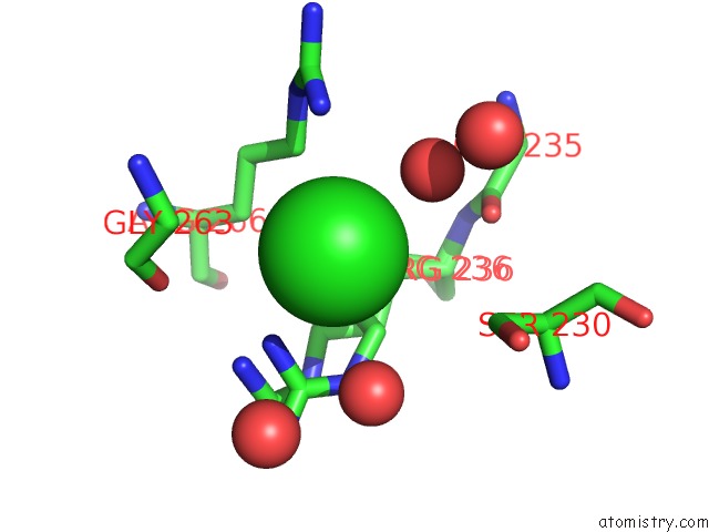
Mono view
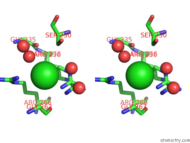
Stereo pair view
|
A full contact list of Chlorine with other atoms in the Cl binding
site number 2 of Crystal Structure of Extended-Spectrum Beta-Lactamase Bel-1 (Orthorhombic Form) within 5.0Å range:
probe
|
atom
|
residue
|
distance (Å)
|
B
|
Occ
|
A:Cl307
b:20.9
occ:1.00
|
NH1
|
A:ARG236
|
3.0
|
12.1
|
0.5
|
O
|
A:HOH506
|
3.1
|
18.3
|
1.0
|
O
|
A:HOH518
|
3.4
|
37.9
|
1.0
|
NH1
|
A:ARG266
|
3.4
|
16.8
|
1.0
|
CD
|
A:ARG236
|
3.5
|
11.3
|
0.5
|
CD
|
A:ARG266
|
3.7
|
13.7
|
1.0
|
CZ
|
A:ARG236
|
3.9
|
12.1
|
0.5
|
CA
|
A:GLY263
|
3.9
|
15.8
|
1.0
|
NH1
|
A:ARG236
|
3.9
|
13.7
|
0.5
|
NE
|
A:ARG236
|
4.0
|
11.9
|
0.5
|
CD
|
A:ARG236
|
4.0
|
12.3
|
0.5
|
CB
|
A:SER230
|
4.2
|
11.7
|
1.0
|
CG
|
A:ARG236
|
4.3
|
10.6
|
0.5
|
CZ
|
A:ARG266
|
4.4
|
15.0
|
1.0
|
CG
|
A:ARG236
|
4.4
|
11.5
|
0.5
|
CB
|
A:ARG236
|
4.5
|
10.9
|
0.5
|
NE
|
A:ARG266
|
4.5
|
14.3
|
1.0
|
O
|
A:GLY263
|
4.5
|
14.7
|
1.0
|
CB
|
A:ARG266
|
4.5
|
11.6
|
1.0
|
O
|
A:HOH408
|
4.6
|
17.3
|
1.0
|
CB
|
A:ARG236
|
4.6
|
10.4
|
0.5
|
CZ
|
A:ARG236
|
4.6
|
13.7
|
0.5
|
CG
|
A:ARG266
|
4.6
|
12.8
|
1.0
|
NE
|
A:ARG236
|
4.7
|
12.9
|
0.5
|
O
|
A:HOH674
|
4.7
|
38.4
|
1.0
|
C
|
A:GLY263
|
4.7
|
15.0
|
1.0
|
O
|
A:GLY235
|
4.7
|
9.8
|
1.0
|
OG
|
A:SER230
|
4.8
|
13.9
|
0.9
|
N
|
A:GLY263
|
4.9
|
15.7
|
1.0
|
NH2
|
A:ARG236
|
4.9
|
12.5
|
0.5
|
|
Chlorine binding site 3 out
of 6 in 5eoe
Go back to
Chlorine Binding Sites List in 5eoe
Chlorine binding site 3 out
of 6 in the Crystal Structure of Extended-Spectrum Beta-Lactamase Bel-1 (Orthorhombic Form)
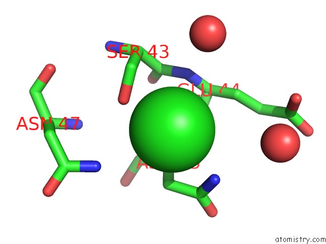
Mono view
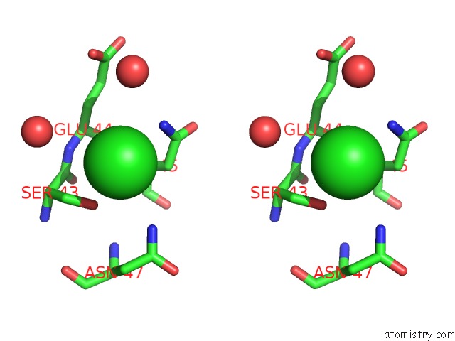
Stereo pair view
|
A full contact list of Chlorine with other atoms in the Cl binding
site number 3 of Crystal Structure of Extended-Spectrum Beta-Lactamase Bel-1 (Orthorhombic Form) within 5.0Å range:
probe
|
atom
|
residue
|
distance (Å)
|
B
|
Occ
|
B:Cl301
b:21.8
occ:1.00
|
OG
|
B:SER43
|
3.0
|
12.7
|
1.0
|
ND2
|
B:ASN45
|
3.1
|
16.1
|
1.0
|
CB
|
B:SER43
|
3.4
|
11.2
|
1.0
|
N
|
B:ASN45
|
3.6
|
11.6
|
1.0
|
CB
|
B:ASN45
|
3.7
|
13.7
|
1.0
|
ND2
|
B:ASN47
|
3.9
|
8.2
|
1.0
|
CG
|
B:ASN45
|
3.9
|
15.6
|
1.0
|
N
|
B:GLU44
|
4.0
|
11.2
|
1.0
|
O
|
B:HOH487
|
4.3
|
18.1
|
1.0
|
CA
|
B:ASN45
|
4.3
|
12.6
|
1.0
|
CB
|
B:GLU44
|
4.3
|
13.3
|
1.0
|
O
|
B:HOH704
|
4.4
|
49.8
|
1.0
|
C
|
B:SER43
|
4.5
|
11.0
|
1.0
|
CA
|
B:GLU44
|
4.5
|
12.3
|
1.0
|
C
|
B:GLU44
|
4.5
|
11.8
|
1.0
|
CA
|
B:SER43
|
4.5
|
11.2
|
1.0
|
CG
|
B:ASN47
|
5.0
|
9.2
|
1.0
|
|
Chlorine binding site 4 out
of 6 in 5eoe
Go back to
Chlorine Binding Sites List in 5eoe
Chlorine binding site 4 out
of 6 in the Crystal Structure of Extended-Spectrum Beta-Lactamase Bel-1 (Orthorhombic Form)
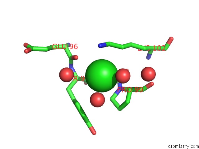
Mono view
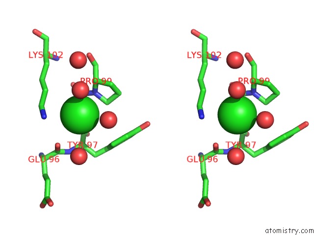
Stereo pair view
|
A full contact list of Chlorine with other atoms in the Cl binding
site number 4 of Crystal Structure of Extended-Spectrum Beta-Lactamase Bel-1 (Orthorhombic Form) within 5.0Å range:
probe
|
atom
|
residue
|
distance (Å)
|
B
|
Occ
|
B:Cl302
b:14.2
occ:0.80
|
O
|
A:HOH727
|
2.9
|
21.2
|
1.0
|
O
|
B:HOH809
|
3.2
|
40.0
|
1.0
|
NZ
|
B:LYS102
|
3.2
|
15.2
|
1.0
|
N
|
B:ALA98
|
3.2
|
8.2
|
1.0
|
N
|
B:PRO99
|
3.5
|
8.8
|
1.0
|
O
|
B:GLU96
|
3.6
|
11.9
|
1.0
|
CD
|
B:PRO99
|
3.7
|
9.3
|
1.0
|
CA
|
B:PRO99
|
3.7
|
8.7
|
1.0
|
C
|
B:ALA98
|
3.7
|
8.4
|
1.0
|
CE
|
B:LYS102
|
3.7
|
13.9
|
1.0
|
O
|
B:HOH619
|
3.8
|
31.8
|
1.0
|
CD
|
B:LYS102
|
3.8
|
11.8
|
1.0
|
CA
|
B:TYR97
|
3.9
|
9.6
|
1.0
|
CB
|
B:PRO99
|
4.0
|
9.0
|
1.0
|
C
|
B:TYR97
|
4.0
|
9.0
|
1.0
|
CA
|
B:ALA98
|
4.1
|
8.4
|
1.0
|
CD1
|
B:TYR97
|
4.2
|
11.5
|
1.0
|
O
|
B:ALA98
|
4.2
|
8.7
|
1.0
|
CG
|
B:PRO99
|
4.4
|
9.6
|
1.0
|
C
|
B:GLU96
|
4.5
|
11.1
|
1.0
|
N
|
B:TYR97
|
4.6
|
10.0
|
1.0
|
CE1
|
B:TYR97
|
4.7
|
12.0
|
1.0
|
CG
|
B:TYR97
|
4.7
|
10.8
|
1.0
|
CB
|
B:TYR97
|
4.8
|
10.5
|
1.0
|
O
|
B:HOH578
|
4.9
|
12.4
|
1.0
|
CG
|
B:LYS102
|
5.0
|
9.8
|
1.0
|
|
Chlorine binding site 5 out
of 6 in 5eoe
Go back to
Chlorine Binding Sites List in 5eoe
Chlorine binding site 5 out
of 6 in the Crystal Structure of Extended-Spectrum Beta-Lactamase Bel-1 (Orthorhombic Form)
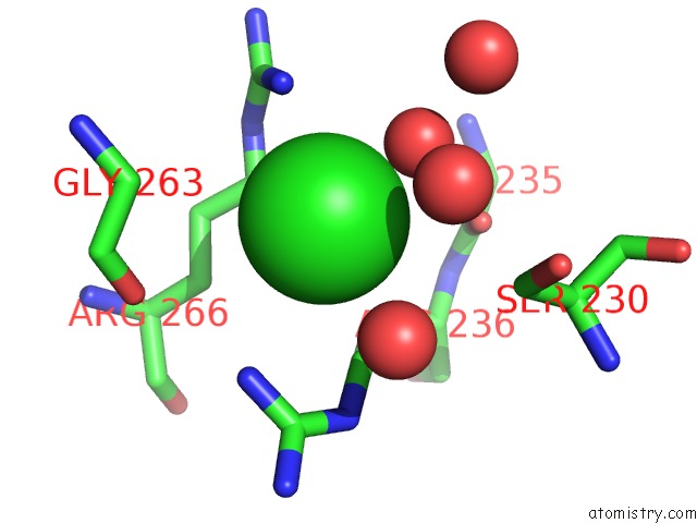
Mono view
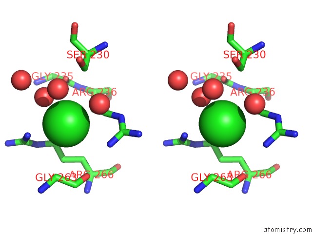
Stereo pair view
|
A full contact list of Chlorine with other atoms in the Cl binding
site number 5 of Crystal Structure of Extended-Spectrum Beta-Lactamase Bel-1 (Orthorhombic Form) within 5.0Å range:
probe
|
atom
|
residue
|
distance (Å)
|
B
|
Occ
|
B:Cl303
b:23.4
occ:1.00
|
O
|
B:HOH489
|
3.1
|
22.1
|
1.0
|
NH1
|
B:ARG266
|
3.3
|
17.6
|
1.0
|
O
|
B:HOH673
|
3.4
|
40.1
|
1.0
|
O
|
B:HOH588
|
3.4
|
44.3
|
1.0
|
CD
|
B:ARG236
|
3.4
|
17.2
|
0.8
|
NH1
|
B:ARG236
|
3.6
|
17.0
|
0.8
|
CD
|
B:ARG266
|
3.7
|
13.6
|
1.0
|
CA
|
B:GLY263
|
4.0
|
14.4
|
1.0
|
NE
|
B:ARG236
|
4.2
|
18.0
|
0.8
|
O
|
B:GLY263
|
4.2
|
14.2
|
1.0
|
CZ
|
B:ARG236
|
4.3
|
18.6
|
0.8
|
CZ
|
B:ARG266
|
4.3
|
16.2
|
1.0
|
CB
|
B:SER230
|
4.3
|
14.3
|
1.0
|
NE
|
B:ARG266
|
4.4
|
15.6
|
1.0
|
CG
|
B:ARG236
|
4.5
|
13.7
|
1.0
|
C
|
B:GLY263
|
4.5
|
15.0
|
1.0
|
CB
|
B:ARG266
|
4.5
|
12.4
|
1.0
|
CG
|
B:ARG266
|
4.5
|
12.3
|
1.0
|
CB
|
B:ARG236
|
4.6
|
11.8
|
1.0
|
O
|
B:HOH409
|
4.6
|
20.7
|
1.0
|
O
|
B:GLY235
|
4.7
|
10.9
|
1.0
|
OG
|
B:SER230
|
4.9
|
15.1
|
0.8
|
|
Chlorine binding site 6 out
of 6 in 5eoe
Go back to
Chlorine Binding Sites List in 5eoe
Chlorine binding site 6 out
of 6 in the Crystal Structure of Extended-Spectrum Beta-Lactamase Bel-1 (Orthorhombic Form)
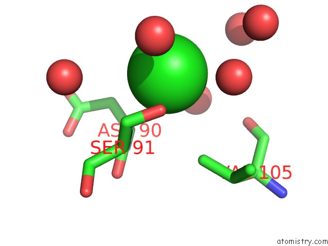
Mono view
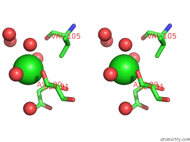
Stereo pair view
|
A full contact list of Chlorine with other atoms in the Cl binding
site number 6 of Crystal Structure of Extended-Spectrum Beta-Lactamase Bel-1 (Orthorhombic Form) within 5.0Å range:
probe
|
atom
|
residue
|
distance (Å)
|
B
|
Occ
|
B:Cl309
b:37.1
occ:1.00
|
O
|
B:HOH720
|
3.1
|
39.0
|
1.0
|
N
|
B:SER91
|
3.1
|
11.5
|
1.0
|
O
|
B:HOH506
|
3.1
|
24.8
|
1.0
|
OG
|
B:SER91
|
3.2
|
17.1
|
1.0
|
CB
|
B:ASP90
|
3.5
|
12.7
|
1.0
|
CA
|
B:ASP90
|
3.6
|
11.3
|
1.0
|
C
|
B:ASP90
|
3.8
|
10.9
|
1.0
|
CB
|
B:SER91
|
3.9
|
13.1
|
1.0
|
CA
|
B:SER91
|
4.0
|
11.4
|
1.0
|
O
|
B:HOH638
|
4.0
|
31.1
|
1.0
|
CG2
|
B:VAL105
|
4.1
|
11.0
|
1.0
|
O
|
B:HOH688
|
4.4
|
37.5
|
1.0
|
O
|
B:HOH653
|
4.4
|
16.5
|
1.0
|
O
|
A:HOH623
|
4.5
|
24.5
|
1.0
|
O
|
B:VAL105
|
4.7
|
9.2
|
1.0
|
CG
|
B:ASP90
|
4.8
|
13.7
|
0.7
|
O
|
B:ASP90
|
5.0
|
10.4
|
1.0
|
N
|
B:ASP90
|
5.0
|
10.6
|
1.0
|
|
Reference:
C.Pozzi,
F.De Luca,
M.Benvenuti,
L.Poirel,
P.Nordmann,
G.M.Rossolini,
S.Mangani,
J.D.Docquier.
Crystal Structure of the Pseudomonas Aeruginosa Bel-1 Extended-Spectrum Beta-Lactamase and Its Complexes with Moxalactam and Imipenem. Antimicrob.Agents Chemother. V. 60 7189 2016.
ISSN: ESSN 1098-6596
PubMed: 27671060
DOI: 10.1128/AAC.00936-16
Page generated: Fri Jul 26 07:25:32 2024
|