Chlorine »
PDB 5eie-5esh »
5eoo »
Chlorine in PDB 5eoo: Crystal Structure of Extended-Spectrum Beta-Lactamase Bel-1 (Monoclinic Form)
Enzymatic activity of Crystal Structure of Extended-Spectrum Beta-Lactamase Bel-1 (Monoclinic Form)
All present enzymatic activity of Crystal Structure of Extended-Spectrum Beta-Lactamase Bel-1 (Monoclinic Form):
3.5.2.6;
3.5.2.6;
Protein crystallography data
The structure of Crystal Structure of Extended-Spectrum Beta-Lactamase Bel-1 (Monoclinic Form), PDB code: 5eoo
was solved by
C.Pozzi,
F.De Luca,
M.Benvenuti,
J.D.Docquier,
S.Mangani,
with X-Ray Crystallography technique. A brief refinement statistics is given in the table below:
| Resolution Low / High (Å) | 51.78 / 1.48 |
| Space group | P 1 21 1 |
| Cell size a, b, c (Å), α, β, γ (°) | 54.840, 94.690, 103.680, 90.00, 92.64, 90.00 |
| R / Rfree (%) | 16.3 / 18.7 |
Chlorine Binding Sites:
The binding sites of Chlorine atom in the Crystal Structure of Extended-Spectrum Beta-Lactamase Bel-1 (Monoclinic Form)
(pdb code 5eoo). This binding sites where shown within
5.0 Angstroms radius around Chlorine atom.
In total 4 binding sites of Chlorine where determined in the Crystal Structure of Extended-Spectrum Beta-Lactamase Bel-1 (Monoclinic Form), PDB code: 5eoo:
Jump to Chlorine binding site number: 1; 2; 3; 4;
In total 4 binding sites of Chlorine where determined in the Crystal Structure of Extended-Spectrum Beta-Lactamase Bel-1 (Monoclinic Form), PDB code: 5eoo:
Jump to Chlorine binding site number: 1; 2; 3; 4;
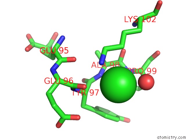
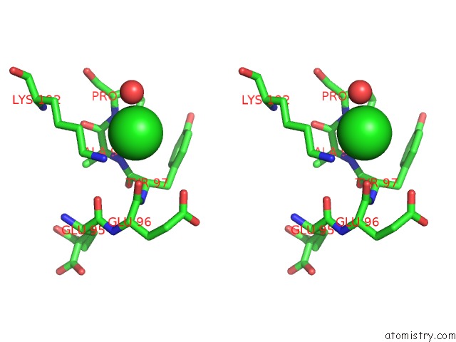
Chlorine binding site 1 out of 4 in 5eoo
Go back to
Chlorine binding site 1 out
of 4 in the Crystal Structure of Extended-Spectrum Beta-Lactamase Bel-1 (Monoclinic Form)
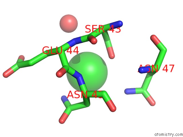
Mono view
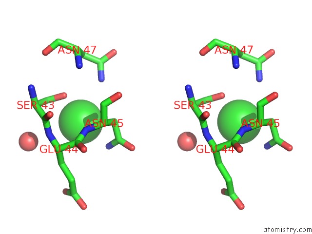
Stereo pair view
Mono view
Stereo pair view
A full contact list of Chlorine with other atoms in the Cl binding
site number 1 of Crystal Structure of Extended-Spectrum Beta-Lactamase Bel-1 (Monoclinic Form) within 5.0Å range:
|
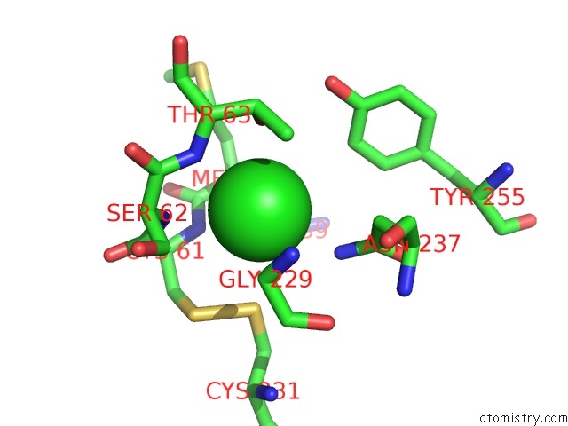
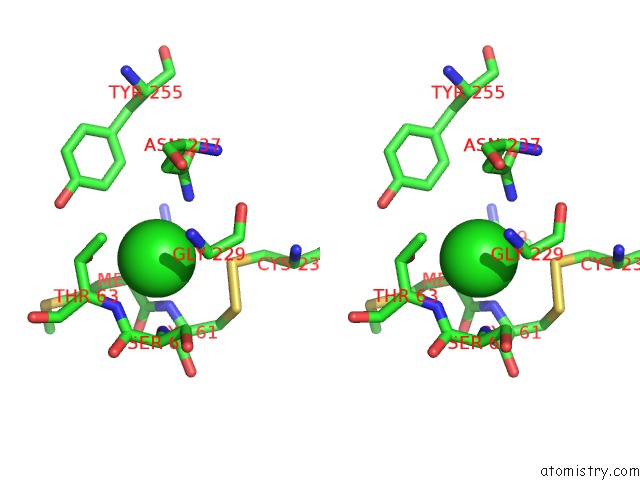
Chlorine binding site 2 out of 4 in 5eoo
Go back to
Chlorine binding site 2 out
of 4 in the Crystal Structure of Extended-Spectrum Beta-Lactamase Bel-1 (Monoclinic Form)
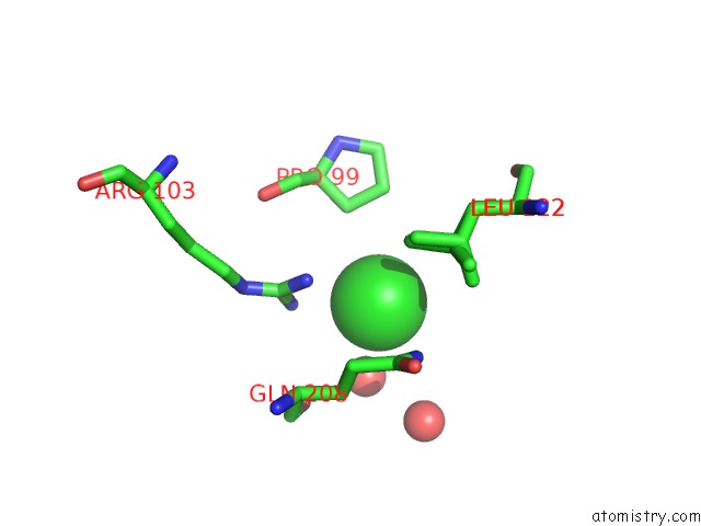
Mono view
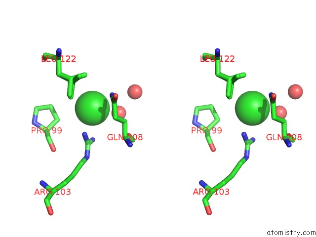
Stereo pair view
Mono view
Stereo pair view
A full contact list of Chlorine with other atoms in the Cl binding
site number 2 of Crystal Structure of Extended-Spectrum Beta-Lactamase Bel-1 (Monoclinic Form) within 5.0Å range:
|
Chlorine binding site 3 out of 4 in 5eoo
Go back to
Chlorine binding site 3 out
of 4 in the Crystal Structure of Extended-Spectrum Beta-Lactamase Bel-1 (Monoclinic Form)
Mono view
Stereo pair view
Mono view
Stereo pair view
A full contact list of Chlorine with other atoms in the Cl binding
site number 3 of Crystal Structure of Extended-Spectrum Beta-Lactamase Bel-1 (Monoclinic Form) within 5.0Å range:
|
Chlorine binding site 4 out of 4 in 5eoo
Go back to
Chlorine binding site 4 out
of 4 in the Crystal Structure of Extended-Spectrum Beta-Lactamase Bel-1 (Monoclinic Form)
Mono view
Stereo pair view
Mono view
Stereo pair view
A full contact list of Chlorine with other atoms in the Cl binding
site number 4 of Crystal Structure of Extended-Spectrum Beta-Lactamase Bel-1 (Monoclinic Form) within 5.0Å range:
|
Reference:
C.Pozzi,
F.De Luca,
M.Benvenuti,
L.Poirel,
P.Nordmann,
G.M.Rossolini,
S.Mangani,
J.D.Docquier.
Crystal Structure of the Pseudomonas Aeruginosa Bel-1 Extended-Spectrum Beta-Lactamase and Its Complexes with Moxalactam and Imipenem. Antimicrob.Agents Chemother. V. 60 7189 2016.
ISSN: ESSN 1098-6596
PubMed: 27671060
DOI: 10.1128/AAC.00936-16
Page generated: Sat Jul 12 01:46:01 2025
ISSN: ESSN 1098-6596
PubMed: 27671060
DOI: 10.1128/AAC.00936-16
Last articles
Fe in 2YXOFe in 2YRS
Fe in 2YXC
Fe in 2YNM
Fe in 2YVJ
Fe in 2YP1
Fe in 2YU2
Fe in 2YU1
Fe in 2YQB
Fe in 2YOO