Chlorine »
PDB 5eie-5esh »
5erk »
Chlorine in PDB 5erk: X-Ray Structure of Horse Spleen Apoferritin (Control)
Protein crystallography data
The structure of X-Ray Structure of Horse Spleen Apoferritin (Control), PDB code: 5erk
was solved by
N.Pontillo,
A.Merlino,
with X-Ray Crystallography technique. A brief refinement statistics is given in the table below:
| Resolution Low / High (Å) | 104.28 / 2.00 |
| Space group | F 4 3 2 |
| Cell size a, b, c (Å), α, β, γ (°) | 180.619, 180.619, 180.619, 90.00, 90.00, 90.00 |
| R / Rfree (%) | 14.3 / 18.1 |
Other elements in 5erk:
The structure of X-Ray Structure of Horse Spleen Apoferritin (Control) also contains other interesting chemical elements:
| Cadmium | (Cd) | 13 atoms |
Chlorine Binding Sites:
The binding sites of Chlorine atom in the X-Ray Structure of Horse Spleen Apoferritin (Control)
(pdb code 5erk). This binding sites where shown within
5.0 Angstroms radius around Chlorine atom.
In total 3 binding sites of Chlorine where determined in the X-Ray Structure of Horse Spleen Apoferritin (Control), PDB code: 5erk:
Jump to Chlorine binding site number: 1; 2; 3;
In total 3 binding sites of Chlorine where determined in the X-Ray Structure of Horse Spleen Apoferritin (Control), PDB code: 5erk:
Jump to Chlorine binding site number: 1; 2; 3;
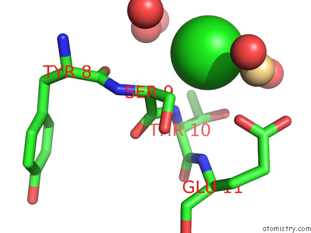
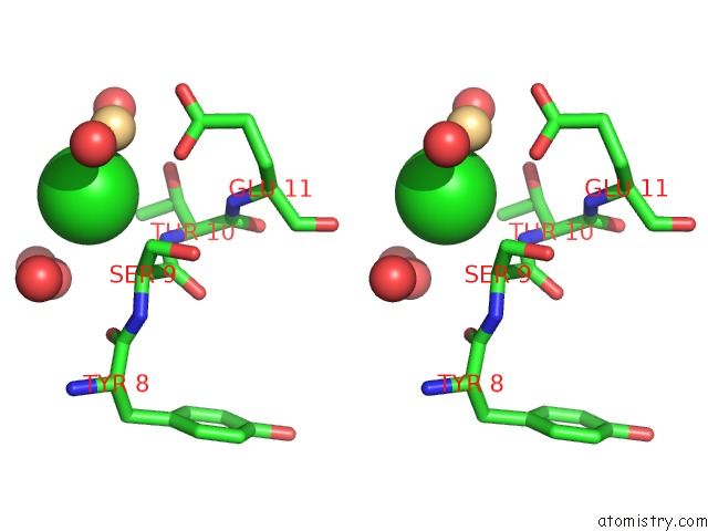
Chlorine binding site 1 out of 3 in 5erk
Go back to
Chlorine binding site 1 out
of 3 in the X-Ray Structure of Horse Spleen Apoferritin (Control)
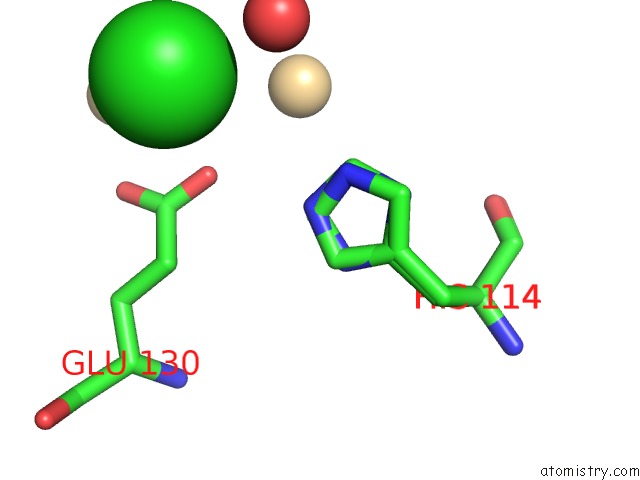
Mono view
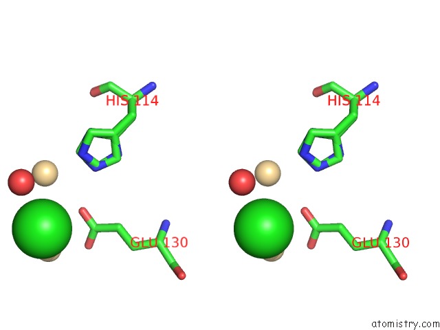
Stereo pair view
Mono view
Stereo pair view
A full contact list of Chlorine with other atoms in the Cl binding
site number 1 of X-Ray Structure of Horse Spleen Apoferritin (Control) within 5.0Å range:
|
Chlorine binding site 2 out of 3 in 5erk
Go back to
Chlorine binding site 2 out
of 3 in the X-Ray Structure of Horse Spleen Apoferritin (Control)
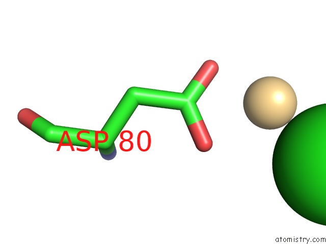
Mono view
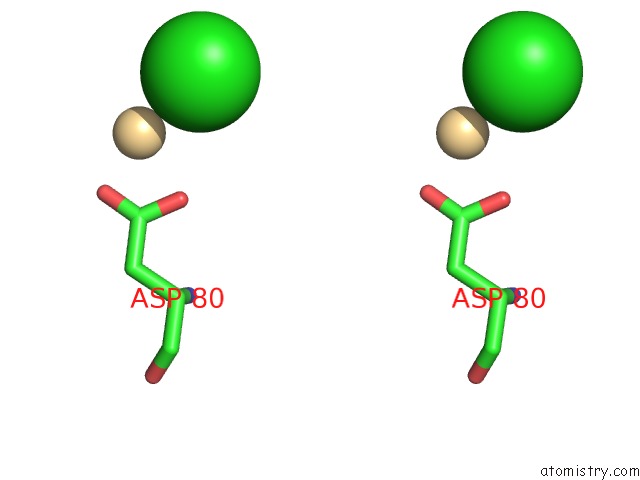
Stereo pair view
Mono view
Stereo pair view
A full contact list of Chlorine with other atoms in the Cl binding
site number 2 of X-Ray Structure of Horse Spleen Apoferritin (Control) within 5.0Å range:
|
Chlorine binding site 3 out of 3 in 5erk
Go back to
Chlorine binding site 3 out
of 3 in the X-Ray Structure of Horse Spleen Apoferritin (Control)
Mono view
Stereo pair view
Mono view
Stereo pair view
A full contact list of Chlorine with other atoms in the Cl binding
site number 3 of X-Ray Structure of Horse Spleen Apoferritin (Control) within 5.0Å range:
|
Reference:
N.Pontillo,
F.Pane,
L.Messori,
A.Amoresano,
A.Merlino.
Cisplatin Encapsulation Within A Ferritin Nanocage: A High-Resolution Crystallographic Study. Chem.Commun.(Camb.) V. 52 4136 2016.
ISSN: ESSN 1364-548X
PubMed: 26888424
DOI: 10.1039/C5CC10365G
Page generated: Sat Jul 12 01:48:20 2025
ISSN: ESSN 1364-548X
PubMed: 26888424
DOI: 10.1039/C5CC10365G
Last articles
Fe in 2YXOFe in 2YRS
Fe in 2YXC
Fe in 2YNM
Fe in 2YVJ
Fe in 2YP1
Fe in 2YU2
Fe in 2YU1
Fe in 2YQB
Fe in 2YOO