Chlorine »
PDB 5f6x-5fdg »
5fbt »
Chlorine in PDB 5fbt: Crystal Structure of Rifampin Phosphotransferase Rph-Lm From Listeria Monocytogenes in Complex with Rifampin
Protein crystallography data
The structure of Crystal Structure of Rifampin Phosphotransferase Rph-Lm From Listeria Monocytogenes in Complex with Rifampin, PDB code: 5fbt
was solved by
P.J.Stogios,
Z.Wawrzak,
T.Skarina,
V.Yim,
A.Savchenko,
W.F.Anderson,
Centerfor Structural Genomics Of Infectious Diseases (Csgid),
with X-Ray Crystallography technique. A brief refinement statistics is given in the table below:
| Resolution Low / High (Å) | 24.98 / 2.70 |
| Space group | P 65 2 2 |
| Cell size a, b, c (Å), α, β, γ (°) | 149.276, 149.276, 193.525, 90.00, 90.00, 120.00 |
| R / Rfree (%) | 22.3 / 26 |
Chlorine Binding Sites:
The binding sites of Chlorine atom in the Crystal Structure of Rifampin Phosphotransferase Rph-Lm From Listeria Monocytogenes in Complex with Rifampin
(pdb code 5fbt). This binding sites where shown within
5.0 Angstroms radius around Chlorine atom.
In total only one binding site of Chlorine was determined in the Crystal Structure of Rifampin Phosphotransferase Rph-Lm From Listeria Monocytogenes in Complex with Rifampin, PDB code: 5fbt:
In total only one binding site of Chlorine was determined in the Crystal Structure of Rifampin Phosphotransferase Rph-Lm From Listeria Monocytogenes in Complex with Rifampin, PDB code: 5fbt:
Chlorine binding site 1 out of 1 in 5fbt
Go back to
Chlorine binding site 1 out
of 1 in the Crystal Structure of Rifampin Phosphotransferase Rph-Lm From Listeria Monocytogenes in Complex with Rifampin
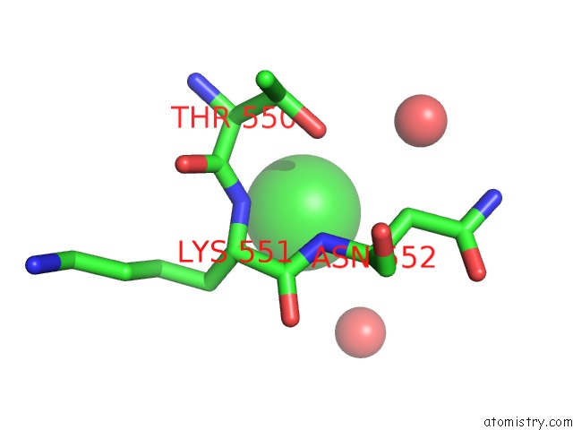
Mono view
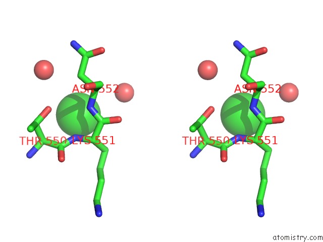
Stereo pair view
Mono view
Stereo pair view
A full contact list of Chlorine with other atoms in the Cl binding
site number 1 of Crystal Structure of Rifampin Phosphotransferase Rph-Lm From Listeria Monocytogenes in Complex with Rifampin within 5.0Å range:
|
Reference:
P.J.Stogios,
G.Cox,
P.Spanogiannopoulos,
M.C.Pillon,
N.Waglechner,
T.Skarina,
K.Koteva,
A.Guarne,
A.Savchenko,
G.D.Wright.
Rifampin Phosphotransferase Is An Unusual Antibiotic Resistance Kinase. Nat Commun V. 7 11343 2016.
ISSN: ESSN 2041-1723
PubMed: 27103605
DOI: 10.1038/NCOMMS11343
Page generated: Sat Jul 12 02:00:06 2025
ISSN: ESSN 2041-1723
PubMed: 27103605
DOI: 10.1038/NCOMMS11343
Last articles
Fe in 2YXOFe in 2YRS
Fe in 2YXC
Fe in 2YNM
Fe in 2YVJ
Fe in 2YP1
Fe in 2YU2
Fe in 2YU1
Fe in 2YQB
Fe in 2YOO