Chlorine »
PDB 5f6x-5fdg »
5fcc »
Chlorine in PDB 5fcc: Structure of Hutd From Pseudomonas Fluorescens SBW25 (Nacl Condition)
Protein crystallography data
The structure of Structure of Hutd From Pseudomonas Fluorescens SBW25 (Nacl Condition), PDB code: 5fcc
was solved by
J.M.Johnston,
M.L.Gerth,
E.N.Baker,
J.S.Lott,
P.B.Rainey,
with X-Ray Crystallography technique. A brief refinement statistics is given in the table below:
| Resolution Low / High (Å) | 54.87 / 1.94 |
| Space group | P 41 21 2 |
| Cell size a, b, c (Å), α, β, γ (°) | 81.998, 81.998, 169.901, 90.00, 90.00, 90.00 |
| R / Rfree (%) | 20 / 23.6 |
Other elements in 5fcc:
The structure of Structure of Hutd From Pseudomonas Fluorescens SBW25 (Nacl Condition) also contains other interesting chemical elements:
| Sodium | (Na) | 1 atom |
Chlorine Binding Sites:
The binding sites of Chlorine atom in the Structure of Hutd From Pseudomonas Fluorescens SBW25 (Nacl Condition)
(pdb code 5fcc). This binding sites where shown within
5.0 Angstroms radius around Chlorine atom.
In total 4 binding sites of Chlorine where determined in the Structure of Hutd From Pseudomonas Fluorescens SBW25 (Nacl Condition), PDB code: 5fcc:
Jump to Chlorine binding site number: 1; 2; 3; 4;
In total 4 binding sites of Chlorine where determined in the Structure of Hutd From Pseudomonas Fluorescens SBW25 (Nacl Condition), PDB code: 5fcc:
Jump to Chlorine binding site number: 1; 2; 3; 4;
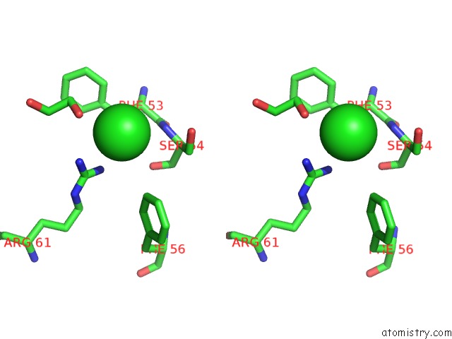
Chlorine binding site 1 out of 4 in 5fcc
Go back to
Chlorine binding site 1 out
of 4 in the Structure of Hutd From Pseudomonas Fluorescens SBW25 (Nacl Condition)

Mono view
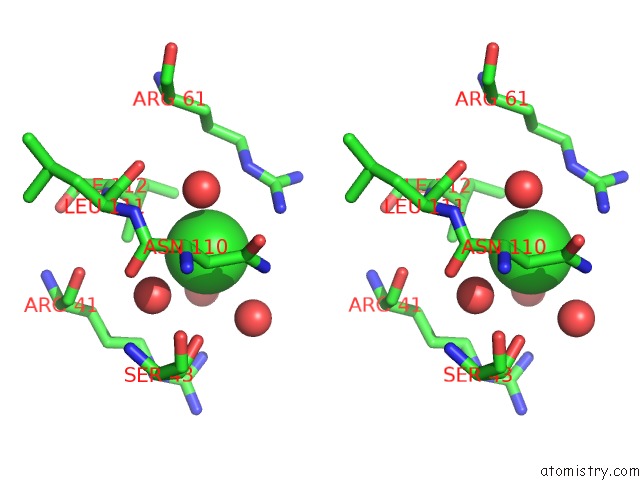
Stereo pair view
Mono view
Stereo pair view
A full contact list of Chlorine with other atoms in the Cl binding
site number 1 of Structure of Hutd From Pseudomonas Fluorescens SBW25 (Nacl Condition) within 5.0Å range:
|
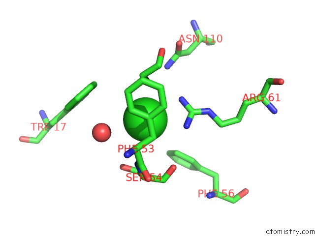
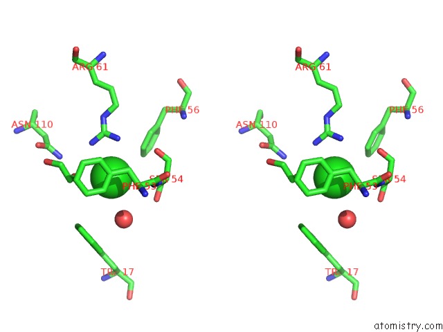
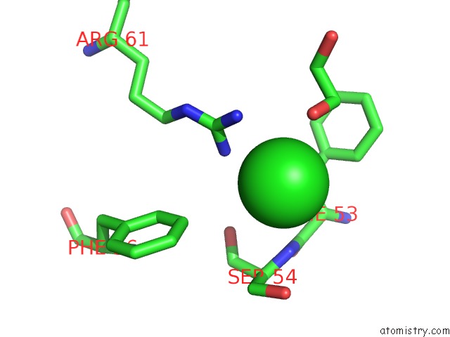
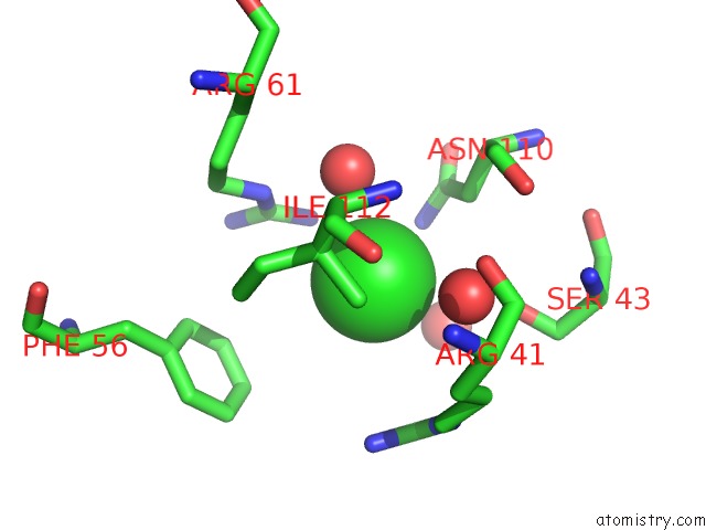
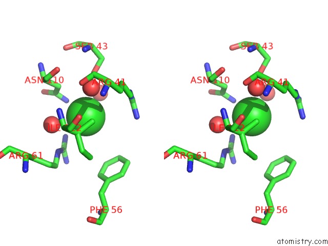
Chlorine binding site 2 out of 4 in 5fcc
Go back to
Chlorine binding site 2 out
of 4 in the Structure of Hutd From Pseudomonas Fluorescens SBW25 (Nacl Condition)
Mono view
Stereo pair view
Mono view
Stereo pair view
A full contact list of Chlorine with other atoms in the Cl binding
site number 2 of Structure of Hutd From Pseudomonas Fluorescens SBW25 (Nacl Condition) within 5.0Å range:
|
Chlorine binding site 3 out of 4 in 5fcc
Go back to
Chlorine binding site 3 out
of 4 in the Structure of Hutd From Pseudomonas Fluorescens SBW25 (Nacl Condition)
Mono view
Stereo pair view
Mono view
Stereo pair view
A full contact list of Chlorine with other atoms in the Cl binding
site number 3 of Structure of Hutd From Pseudomonas Fluorescens SBW25 (Nacl Condition) within 5.0Å range:
|
Chlorine binding site 4 out of 4 in 5fcc
Go back to
Chlorine binding site 4 out
of 4 in the Structure of Hutd From Pseudomonas Fluorescens SBW25 (Nacl Condition)
Mono view
Stereo pair view
Mono view
Stereo pair view
A full contact list of Chlorine with other atoms in the Cl binding
site number 4 of Structure of Hutd From Pseudomonas Fluorescens SBW25 (Nacl Condition) within 5.0Å range:
|
Reference:
M.L.Gerth,
Y.H.Liu,
X.-X.Zhang,
E.N.Baker,
J.S.Lott,
P.B.Rainey,
J.M.Johnston.
Structure of Hutd From Pseudomonas Fluorescens To Be Published.
Page generated: Sat Jul 12 02:00:29 2025
Last articles
Fe in 2YXOFe in 2YRS
Fe in 2YXC
Fe in 2YNM
Fe in 2YVJ
Fe in 2YP1
Fe in 2YU2
Fe in 2YU1
Fe in 2YQB
Fe in 2YOO