Chlorine »
PDB 5fzb-5g60 »
5g36 »
Chlorine in PDB 5g36: Yellow Form of Halorhodopsin From Halobacterium Salinarum in A New Rhombohedral Crystal Form
Protein crystallography data
The structure of Yellow Form of Halorhodopsin From Halobacterium Salinarum in A New Rhombohedral Crystal Form, PDB code: 5g36
was solved by
M.Schreiner,
R.Schlesinger,
J.Heberle,
H.H.Niemann,
with X-Ray Crystallography technique. A brief refinement statistics is given in the table below:
| Resolution Low / High (Å) | 42.39 / 2.60 |
| Space group | H 3 2 |
| Cell size a, b, c (Å), α, β, γ (°) | 103.000, 103.000, 136.450, 90.00, 90.00, 120.00 |
| R / Rfree (%) | 19.1 / 25.3 |
Chlorine Binding Sites:
The binding sites of Chlorine atom in the Yellow Form of Halorhodopsin From Halobacterium Salinarum in A New Rhombohedral Crystal Form
(pdb code 5g36). This binding sites where shown within
5.0 Angstroms radius around Chlorine atom.
In total only one binding site of Chlorine was determined in the Yellow Form of Halorhodopsin From Halobacterium Salinarum in A New Rhombohedral Crystal Form, PDB code: 5g36:
In total only one binding site of Chlorine was determined in the Yellow Form of Halorhodopsin From Halobacterium Salinarum in A New Rhombohedral Crystal Form, PDB code: 5g36:
Chlorine binding site 1 out of 1 in 5g36
Go back to
Chlorine binding site 1 out
of 1 in the Yellow Form of Halorhodopsin From Halobacterium Salinarum in A New Rhombohedral Crystal Form
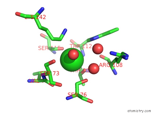
Mono view
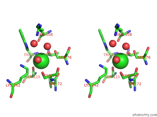
Stereo pair view
Mono view
Stereo pair view
A full contact list of Chlorine with other atoms in the Cl binding
site number 1 of Yellow Form of Halorhodopsin From Halobacterium Salinarum in A New Rhombohedral Crystal Form within 5.0Å range:
|
Reference:
M.Schreiner,
R.Schlesinger,
J.Heberle,
H.H.Niemann.
Crystal Structure of Halobacterium Salinarum Halorhodopsin with Partially Depopulated Primary Chloride Binding Site Acta Crystallogr.,Sect.F V. 72 692 2016.
ISSN: ESSN 1744-3091
PubMed: 27599860
DOI: 10.1107/S2053230X16012796
Page generated: Fri Jul 26 08:19:19 2024
ISSN: ESSN 1744-3091
PubMed: 27599860
DOI: 10.1107/S2053230X16012796
Last articles
Zn in 9J0NZn in 9J0O
Zn in 9J0P
Zn in 9FJX
Zn in 9EKB
Zn in 9C0F
Zn in 9CAH
Zn in 9CH0
Zn in 9CH3
Zn in 9CH1