Atomistry » Chlorine » PDB 5fz9-5g5n » 5g3t
Atomistry »
  Chlorine »
    PDB 5fz9-5g5n »
      5g3t »

Chlorine in PDB 5g3t: The Structure of the L-Tryptophan Oxidase Vioa From Chromobacterium Violaceum

Enzymatic activity of The Structure of the L-Tryptophan Oxidase Vioa From Chromobacterium Violaceum

All present enzymatic activity of The Structure of the L-Tryptophan Oxidase Vioa From Chromobacterium Violaceum:
1.4.3.23;

Protein crystallography data

The structure of The Structure of the L-Tryptophan Oxidase Vioa From Chromobacterium Violaceum, PDB code: 5g3t was solved by J.Krausze, J.Rabe, J.Moser, with X-Ray Crystallography technique. A brief refinement statistics is given in the table below:

Resolution Low / High (Å) 19.99 / 1.80
Space group P 1 21 1
Cell size a, b, c (Å), α, β, γ (°) 68.090, 89.160, 144.430, 90.00, 92.66, 90.00
R / Rfree (%) 15.7 / 19.2

Other elements in 5g3t:

The structure of The Structure of the L-Tryptophan Oxidase Vioa From Chromobacterium Violaceum also contains other interesting chemical elements:

Magnesium (Mg) 6 atoms

Chlorine Binding Sites:

Pages:

>>> Page 1 <<< Page 2, Binding sites: 11 - 11;

Binding sites:

The binding sites of Chlorine atom in the The Structure of the L-Tryptophan Oxidase Vioa From Chromobacterium Violaceum (pdb code 5g3t). This binding sites where shown within 5.0 Angstroms radius around Chlorine atom.
In total 11 binding sites of Chlorine where determined in the The Structure of the L-Tryptophan Oxidase Vioa From Chromobacterium Violaceum, PDB code: 5g3t:
Jump to Chlorine binding site number: 1; 2; 3; 4; 5; 6; 7; 8; 9; 10;

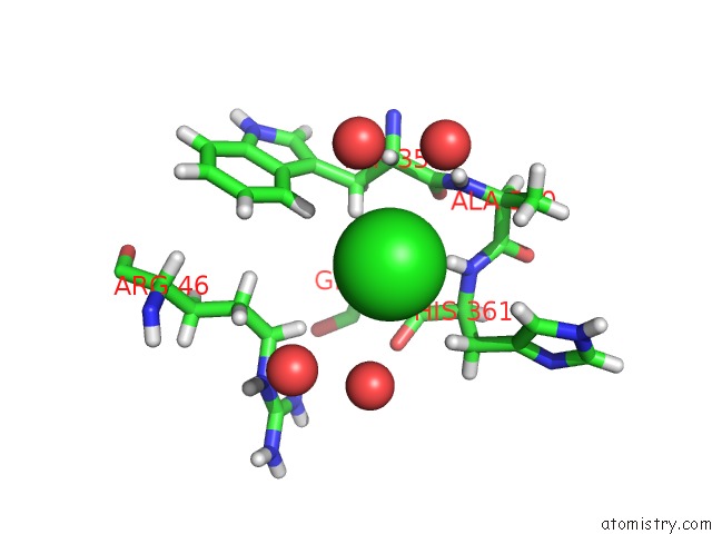
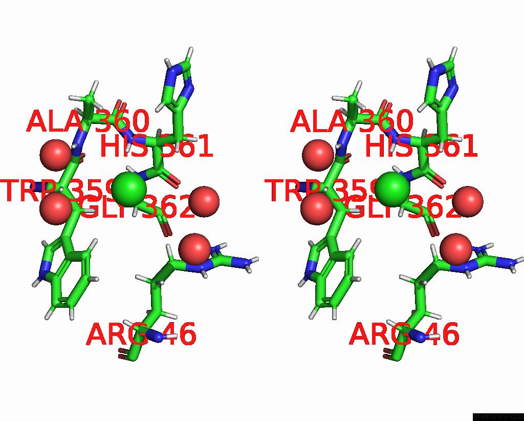
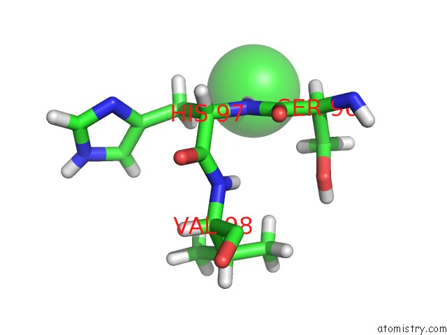
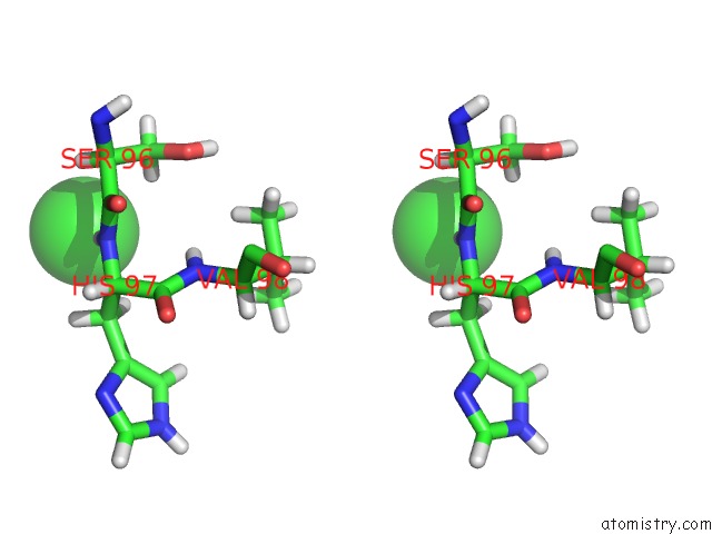
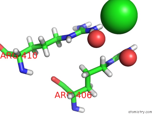
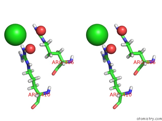
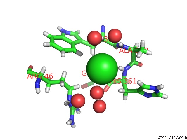
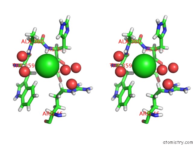
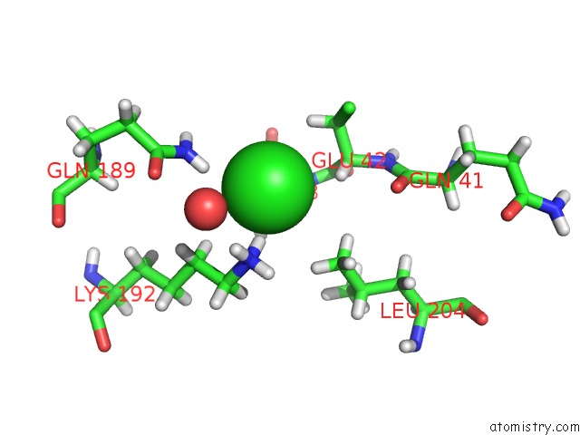
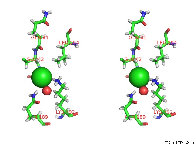
Chlorine binding site 1 out of 11 in 5g3t

Go back to Chlorine Binding Sites List in 5g3t
Chlorine binding site 1 out of 11 in the The Structure of the L-Tryptophan Oxidase Vioa From Chromobacterium Violaceum


Mono view


Stereo pair view

A full contact list of Chlorine with other atoms in the Cl binding site number 1 of The Structure of the L-Tryptophan Oxidase Vioa From Chromobacterium Violaceum within 5.0Å range:
probe atom residue distance (Å) B Occ
A:Cl421

b:23.1
occ:1.00
H A:HIS361 2.3 19.6 1.0
HB3 A:TRP359 2.9 14.0 1.0
HE3 A:TRP359 3.0 16.4 1.0
N A:HIS361 3.1 16.3 1.0
HB2 A:HIS361 3.1 26.4 1.0
C A:HIS361 3.4 13.1 1.0
HA A:TRP359 3.5 16.8 1.0
H A:ALA360 3.5 14.3 1.0
HA2 A:GLY362 3.6 13.8 1.0
N A:ALA360 3.6 11.9 1.0
CA A:HIS361 3.6 16.0 1.0
O A:HIS361 3.6 18.8 1.0
HD3 A:ARG46 3.7 19.8 1.0
CB A:TRP359 3.8 11.7 1.0
C A:TRP359 3.8 13.2 1.0
N A:GLY362 3.8 11.6 1.0
O A:HOH2051 3.8 24.8 1.0
CB A:HIS361 3.8 22.0 1.0
CA A:TRP359 3.8 14.0 1.0
CE3 A:TRP359 3.9 13.7 1.0
H A:GLY362 4.2 13.9 1.0
O A:HOH2326 4.2 21.8 1.0
C A:ALA360 4.2 15.2 1.0
CA A:GLY362 4.2 11.5 1.0
HB1 A:ALA360 4.2 25.5 1.0
HD2 A:HIS361 4.2 26.2 1.0
HB2 A:TRP359 4.4 14.0 1.0
CA A:ALA360 4.4 13.8 1.0
O A:TRP359 4.4 13.9 1.0
HG2 A:ARG46 4.5 19.6 1.0
HB3 A:HIS361 4.5 26.4 1.0
HA A:HIS361 4.6 19.2 1.0
CD2 A:TRP359 4.6 11.8 1.0
CG A:TRP359 4.6 9.8 1.0
CD A:ARG46 4.6 16.5 1.0
CG A:HIS361 4.7 23.7 1.0
HA3 A:GLY362 4.8 13.8 1.0
CD2 A:HIS361 4.8 21.9 1.0
O A:HOH2324 4.8 33.9 1.0
CB A:ALA360 4.9 21.2 1.0
HG3 A:ARG46 4.9 19.6 1.0
HZ3 A:TRP359 4.9 17.9 1.0
CG A:ARG46 4.9 16.3 1.0
CZ3 A:TRP359 4.9 14.9 1.0
O A:HOH2357 4.9 21.0 1.0

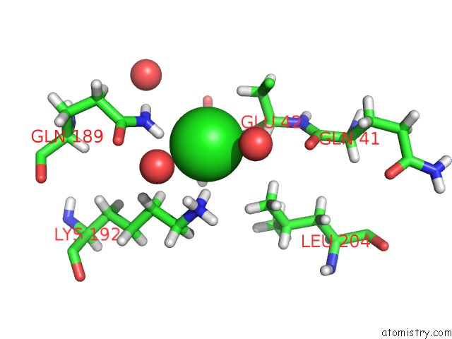
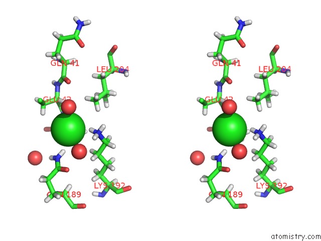
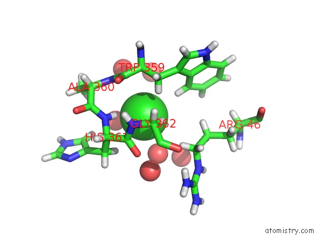
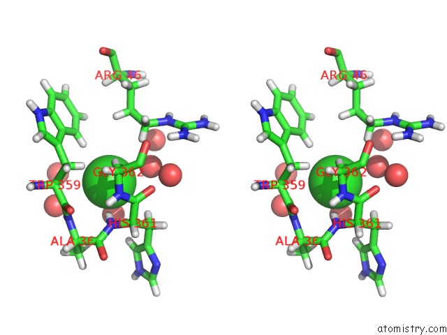
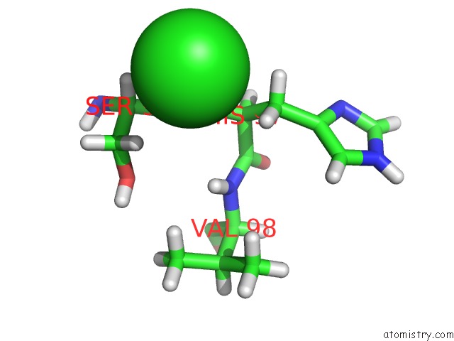
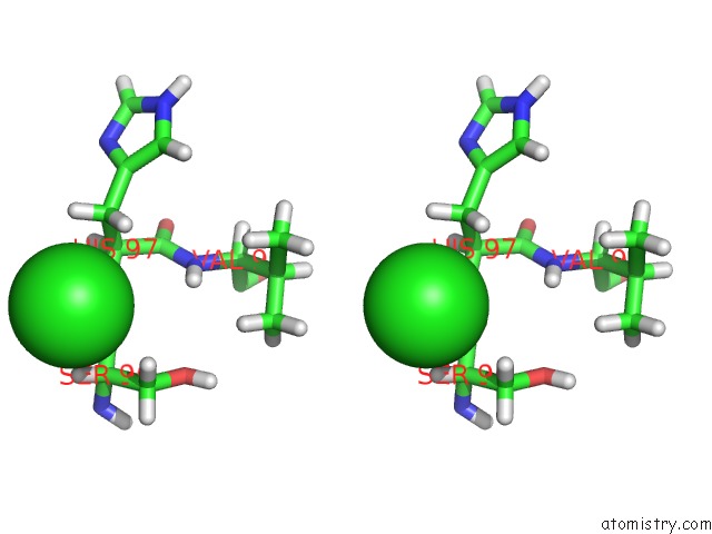
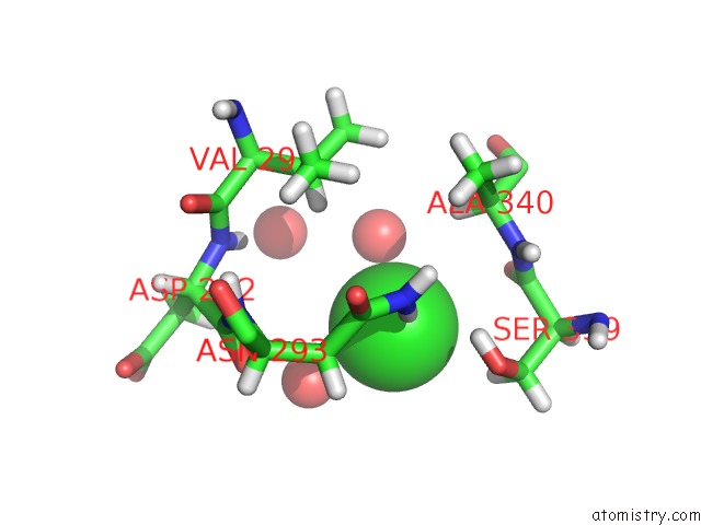
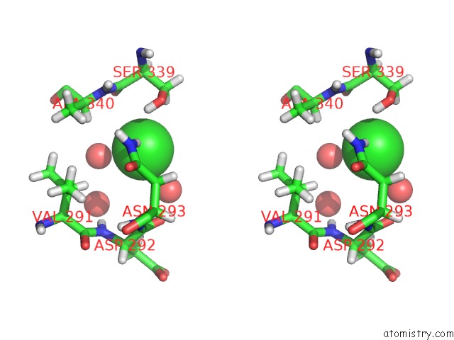
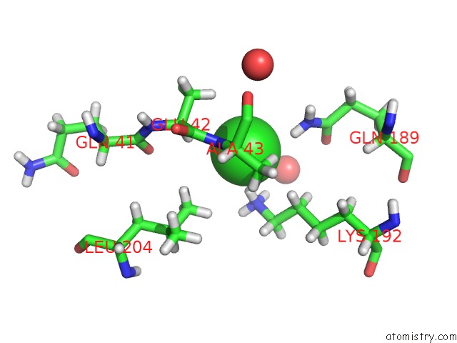
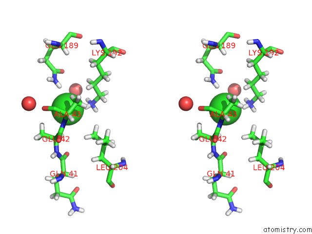
Chlorine binding site 2 out of 11 in 5g3t

Go back to Chlorine Binding Sites List in 5g3t
Chlorine binding site 2 out of 11 in the The Structure of the L-Tryptophan Oxidase Vioa From Chromobacterium Violaceum


Mono view


Stereo pair view

A full contact list of Chlorine with other atoms in the Cl binding site number 2 of The Structure of the L-Tryptophan Oxidase Vioa From Chromobacterium Violaceum within 5.0Å range:
probe atom residue distance (Å) B Occ
A:Cl422

b:28.5
occ:1.00
H A:ALA43 2.3 28.0 1.0
HE22 A:GLN189 2.3 30.5 1.0
HZ3 A:LYS192 2.4 34.0 1.0
O A:HOH2040 2.9 30.0 1.0
HA A:GLU42 3.0 25.8 1.0
NE2 A:GLN189 3.1 25.4 1.0
N A:ALA43 3.2 23.3 1.0
NZ A:LYS192 3.2 28.3 1.0
HB3 A:GLU42 3.3 40.2 1.0
HZ1 A:LYS192 3.3 34.0 1.0
HD2 A:LYS192 3.4 28.6 1.0
HB1 A:ALA43 3.5 20.4 1.0
HE21 A:GLN189 3.5 30.5 1.0
CA A:GLU42 3.6 21.5 1.0
HD12 A:LEU204 3.7 39.4 1.0
CB A:GLU42 3.8 33.5 1.0
HD11 A:LEU204 3.8 39.4 1.0
HZ2 A:LYS192 3.8 34.0 1.0
O A:HOH2225 3.9 40.8 1.0
HD3 A:LYS192 3.9 28.6 1.0
C A:GLU42 3.9 22.0 1.0
HB3 A:ALA43 3.9 20.4 1.0
CD A:LYS192 3.9 23.8 1.0
CB A:ALA43 4.0 17.0 1.0
CG A:GLU42 4.0 36.1 1.0
CE A:LYS192 4.0 23.4 1.0
CD A:GLN189 4.1 40.6 1.0
CD1 A:LEU204 4.1 32.8 1.0
CA A:ALA43 4.2 16.8 1.0
OE1 A:GLN189 4.2 34.0 1.0
HE3 A:LYS192 4.3 28.1 1.0
HD13 A:LEU204 4.3 39.4 1.0
HB2 A:GLU42 4.7 40.2 1.0
O A:ALA43 4.7 18.7 1.0
O A:GLN41 4.8 27.9 1.0
O A:HOH2222 4.8 44.7 1.0
HE2 A:LYS192 4.9 28.1 1.0
C A:ALA43 4.9 16.7 1.0
HA A:ALA43 4.9 20.2 1.0
HB2 A:ALA43 4.9 20.4 1.0
N A:GLU42 4.9 26.3 1.0

Chlorine binding site 3 out of 11 in 5g3t

Go back to Chlorine Binding Sites List in 5g3t
Chlorine binding site 3 out of 11 in the The Structure of the L-Tryptophan Oxidase Vioa From Chromobacterium Violaceum


Mono view


Stereo pair view

A full contact list of Chlorine with other atoms in the Cl binding site number 3 of The Structure of the L-Tryptophan Oxidase Vioa From Chromobacterium Violaceum within 5.0Å range:
probe atom residue distance (Å) B Occ
B:Cl422

b:23.4
occ:1.00
H B:HIS97 2.4 18.9 1.0
HB3 B:HIS97 3.1 18.6 1.0
HB2 B:SER96 3.2 24.1 1.0
N B:HIS97 3.2 15.7 1.0
HA B:SER96 3.3 19.3 1.0
HB2 B:HIS97 3.5 18.6 1.0
CB B:HIS97 3.7 15.5 1.0
CA B:SER96 3.9 16.1 1.0
CB B:SER96 3.9 20.1 1.0
C B:SER96 4.1 16.1 1.0
CA B:HIS97 4.1 12.8 1.0
HB3 B:SER96 4.3 24.1 1.0
H B:VAL98 4.5 14.6 1.0
HA B:HIS97 4.7 15.3 1.0

Chlorine binding site 4 out of 11 in 5g3t

Go back to Chlorine Binding Sites List in 5g3t
Chlorine binding site 4 out of 11 in the The Structure of the L-Tryptophan Oxidase Vioa From Chromobacterium Violaceum


Mono view


Stereo pair view

A full contact list of Chlorine with other atoms in the Cl binding site number 4 of The Structure of the L-Tryptophan Oxidase Vioa From Chromobacterium Violaceum within 5.0Å range:
probe atom residue distance (Å) B Occ
B:Cl423

b:25.3
occ:1.00
H B:HIS361 2.4 21.9 1.0
HB3 B:TRP359 2.9 16.6 1.0
HE3 B:TRP359 3.0 19.3 1.0
O A:HOH2108 3.1 39.0 1.0
O B:HOH2008 3.1 37.0 1.0
N B:HIS361 3.2 18.3 1.0
HB2 B:HIS361 3.3 30.8 1.0
C B:HIS361 3.4 20.6 1.0
H B:ALA360 3.5 17.7 1.0
HA B:TRP359 3.5 18.4 1.0
HA2 B:GLY362 3.6 18.1 1.0
N B:ALA360 3.6 14.7 1.0
CA B:HIS361 3.7 20.3 1.0
HD3 B:ARG46 3.7 23.7 1.0
O B:HIS361 3.7 20.7 1.0
CB B:TRP359 3.7 13.9 1.0
N B:GLY362 3.8 14.9 1.0
C B:TRP359 3.8 16.9 1.0
CA B:TRP359 3.8 15.3 1.0
CE3 B:TRP359 3.9 16.1 1.0
CB B:HIS361 3.9 25.6 1.0
O B:HOH2043 4.0 26.0 1.0
H B:GLY362 4.1 17.8 1.0
CA B:GLY362 4.2 15.1 1.0
O A:HOH2251 4.2 24.4 1.0
C B:ALA360 4.2 18.3 1.0
HD2 B:HIS361 4.3 31.1 1.0
HG2 B:ARG46 4.3 23.7 1.0
HB1 B:ALA360 4.3 30.2 1.0
HB2 B:TRP359 4.4 16.6 1.0
O B:TRP359 4.4 17.6 1.0
CA B:ALA360 4.4 17.3 1.0
CD B:ARG46 4.6 19.7 1.0
CD2 B:TRP359 4.6 14.6 1.0
CG B:TRP359 4.6 12.7 1.0
HA B:HIS361 4.6 24.4 1.0
HB3 B:HIS361 4.6 30.8 1.0
O B:HOH2282 4.7 42.3 1.0
CG B:HIS361 4.8 28.4 1.0
HA3 B:GLY362 4.8 18.1 1.0
HG3 B:ARG46 4.8 23.7 1.0
CG B:ARG46 4.8 19.8 1.0
CD2 B:HIS361 4.8 25.9 1.0
O B:HOH2299 4.9 25.7 1.0
HZ3 B:TRP359 4.9 19.3 1.0
CZ3 B:TRP359 4.9 16.1 1.0
CB B:ALA360 4.9 25.2 1.0

Chlorine binding site 5 out of 11 in 5g3t

Go back to Chlorine Binding Sites List in 5g3t
Chlorine binding site 5 out of 11 in the The Structure of the L-Tryptophan Oxidase Vioa From Chromobacterium Violaceum


Mono view


Stereo pair view

A full contact list of Chlorine with other atoms in the Cl binding site number 5 of The Structure of the L-Tryptophan Oxidase Vioa From Chromobacterium Violaceum within 5.0Å range:
probe atom residue distance (Å) B Occ
C:Cl420

b:57.1
occ:1.00
HH22 C:ARG410 2.2 36.4 1.0
NH2 C:ARG410 3.0 30.4 1.0
HH12 C:ARG410 3.2 31.7 1.0
HH12 C:ARG406 3.3 49.9 1.0
NH1 C:ARG406 3.5 41.6 1.0
HH21 C:ARG410 3.5 36.4 1.0
HH11 C:ARG406 3.7 49.9 1.0
NH1 C:ARG410 3.8 26.4 1.0
CZ C:ARG410 3.9 22.5 1.0
CZ C:ARG406 4.0 39.5 1.0
HH22 C:ARG406 4.1 41.1 1.0
NH2 C:ARG406 4.3 34.3 1.0
HD2 C:ARG406 4.5 30.8 1.0
HH11 C:ARG410 4.6 31.7 1.0
O C:HOH2347 4.7 35.9 1.0
NE C:ARG406 4.7 33.4 1.0
HH21 C:ARG406 4.8 41.1 1.0
O C:HOH2343 4.9 37.6 1.0

Chlorine binding site 6 out of 11 in 5g3t

Go back to Chlorine Binding Sites List in 5g3t
Chlorine binding site 6 out of 11 in the The Structure of the L-Tryptophan Oxidase Vioa From Chromobacterium Violaceum


Mono view


Stereo pair view

A full contact list of Chlorine with other atoms in the Cl binding site number 6 of The Structure of the L-Tryptophan Oxidase Vioa From Chromobacterium Violaceum within 5.0Å range:
probe atom residue distance (Å) B Occ
C:Cl421

b:25.5
occ:1.00
H C:HIS97 2.3 22.9 1.0
HB3 C:HIS97 3.2 18.3 1.0
N C:HIS97 3.2 19.1 1.0
HB2 C:SER96 3.2 22.0 1.0
HA C:SER96 3.4 23.6 1.0
HB2 C:HIS97 3.5 18.3 1.0
CB C:HIS97 3.7 15.3 1.0
CA C:SER96 3.9 19.7 1.0
CB C:SER96 4.0 18.3 1.0
C C:SER96 4.1 21.8 1.0
CA C:HIS97 4.1 16.9 1.0
HB3 C:SER96 4.4 22.0 1.0
H C:VAL98 4.5 17.0 1.0
HA C:HIS97 4.7 20.3 1.0

Chlorine binding site 7 out of 11 in 5g3t

Go back to Chlorine Binding Sites List in 5g3t
Chlorine binding site 7 out of 11 in the The Structure of the L-Tryptophan Oxidase Vioa From Chromobacterium Violaceum


Mono view


Stereo pair view

A full contact list of Chlorine with other atoms in the Cl binding site number 7 of The Structure of the L-Tryptophan Oxidase Vioa From Chromobacterium Violaceum within 5.0Å range:
probe atom residue distance (Å) B Occ
C:Cl422

b:24.2
occ:1.00
H C:HIS361 2.3 18.2 1.0
O C:HOH2020 2.8 30.9 1.0
HE3 C:TRP359 2.9 16.9 1.0
HB3 C:TRP359 3.0 13.5 1.0
N C:HIS361 3.2 15.2 1.0
HB2 C:HIS361 3.2 24.8 1.0
H C:ALA360 3.4 15.4 1.0
C C:HIS361 3.4 14.7 1.0
HA C:TRP359 3.5 12.9 1.0
HA2 C:GLY362 3.6 17.8 1.0
N C:ALA360 3.6 12.8 1.0
O C:HIS361 3.6 18.1 1.0
HD3 C:ARG46 3.6 20.2 1.0
CA C:HIS361 3.7 16.1 1.0
N C:GLY362 3.8 13.0 1.0
C C:TRP359 3.8 13.7 1.0
CB C:TRP359 3.8 11.3 1.0
CE3 C:TRP359 3.8 14.1 1.0
CA C:TRP359 3.9 10.8 1.0
CB C:HIS361 3.9 20.7 1.0
O C:HOH2328 4.1 20.6 1.0
H C:GLY362 4.2 15.7 1.0
CA C:GLY362 4.2 14.9 1.0
C C:ALA360 4.2 15.7 1.0
HB1 C:ALA360 4.3 19.8 1.0
O C:HOH2057 4.3 25.5 1.0
HG2 C:ARG46 4.3 16.1 1.0
HD2 C:HIS361 4.4 28.2 1.0
CA C:ALA360 4.4 13.8 1.0
HB2 C:TRP359 4.4 13.5 1.0
O C:TRP359 4.5 13.7 1.0
CD C:ARG46 4.5 16.8 1.0
CD2 C:TRP359 4.5 10.9 1.0
HA C:HIS361 4.6 19.4 1.0
HB3 C:HIS361 4.6 24.8 1.0
CG C:TRP359 4.6 10.4 1.0
O C:HOH2326 4.7 30.0 1.0
HG3 C:ARG46 4.7 16.1 1.0
HZ3 C:TRP359 4.7 16.4 1.0
CG C:ARG46 4.7 13.4 1.0
HA3 C:GLY362 4.7 17.8 1.0
CZ3 C:TRP359 4.8 13.7 1.0
CG C:HIS361 4.8 19.5 1.0
CB C:ALA360 4.9 16.5 1.0
O C:HOH2355 4.9 22.1 1.0
CD2 C:HIS361 4.9 23.5 1.0

Chlorine binding site 8 out of 11 in 5g3t

Go back to Chlorine Binding Sites List in 5g3t
Chlorine binding site 8 out of 11 in the The Structure of the L-Tryptophan Oxidase Vioa From Chromobacterium Violaceum


Mono view


Stereo pair view

A full contact list of Chlorine with other atoms in the Cl binding site number 8 of The Structure of the L-Tryptophan Oxidase Vioa From Chromobacterium Violaceum within 5.0Å range:
probe atom residue distance (Å) B Occ
C:Cl423

b:26.2
occ:1.00
HD21 C:ASN293 2.5 20.6 1.0
O C:HOH2147 2.9 33.4 1.0
HG C:SER339 2.9 31.5 1.0
HB3 C:ASN293 2.9 24.5 1.0
O C:HOH2283 3.0 16.3 1.0
HB2 C:SER339 3.0 21.7 1.0
ND2 C:ASN293 3.3 17.2 1.0
OG C:SER339 3.4 26.3 1.0
HA C:ALA340 3.6 22.9 1.0
CB C:SER339 3.7 18.1 1.0
O C:SER339 3.8 21.1 1.0
C C:SER339 3.8 23.0 1.0
CB C:ASN293 3.8 20.4 1.0
HD22 C:ASN293 3.8 20.6 1.0
HB C:VAL291 4.0 19.6 1.0
CG C:ASN293 4.1 17.5 1.0
N C:ALA340 4.1 21.1 1.0
HB2 C:ASN293 4.1 24.5 1.0
O C:ASP292 4.2 11.8 1.0
CA C:ALA340 4.3 19.1 1.0
CA C:SER339 4.4 19.0 1.0
HB3 C:SER339 4.5 21.7 1.0
H C:ALA340 4.5 25.2 1.0
O C:HOH2282 4.5 20.9 1.0
HB3 C:ALA340 4.6 25.5 1.0
C C:ASP292 4.6 12.3 1.0
H C:ASP292 4.8 15.7 1.0
HA C:SER339 4.8 22.8 1.0
N C:ASN293 4.9 13.1 1.0
CA C:ASN293 4.9 11.8 1.0
CB C:VAL291 5.0 16.3 1.0

Chlorine binding site 9 out of 11 in 5g3t

Go back to Chlorine Binding Sites List in 5g3t
Chlorine binding site 9 out of 11 in the The Structure of the L-Tryptophan Oxidase Vioa From Chromobacterium Violaceum


Mono view


Stereo pair view

A full contact list of Chlorine with other atoms in the Cl binding site number 9 of The Structure of the L-Tryptophan Oxidase Vioa From Chromobacterium Violaceum within 5.0Å range:
probe atom residue distance (Å) B Occ
C:Cl424

b:25.8
occ:1.00
HZ3 C:LYS192 2.2 32.2 1.0
H C:ALA43 2.3 21.8 1.0
HE22 C:GLN189 2.6 27.9 1.0
HA C:GLU42 2.9 25.3 1.0
NZ C:LYS192 3.0 26.9 1.0
N C:ALA43 3.1 18.1 1.0
HZ1 C:LYS192 3.2 32.2 1.0
HB3 C:GLU42 3.4 32.4 1.0
HD2 C:LYS192 3.4 24.4 1.0
HB1 C:ALA43 3.4 23.2 1.0
NE2 C:GLN189 3.4 23.2 1.0
HZ2 C:LYS192 3.5 32.2 1.0
HD12 C:LEU204 3.6 29.6 1.0
CA C:GLU42 3.6 21.1 1.0
HD11 C:LEU204 3.7 29.6 1.0
O C:HOH2216 3.8 36.6 1.0
HE21 C:GLN189 3.8 27.9 1.0
HB3 C:ALA43 3.8 23.2 1.0
CB C:GLU42 3.9 27.0 1.0
C C:GLU42 3.9 21.7 1.0
CE C:LYS192 3.9 24.9 1.0
CD C:LYS192 3.9 20.4 1.0
CB C:ALA43 3.9 19.4 1.0
HD3 C:LYS192 4.0 24.4 1.0
CD1 C:LEU204 4.0 24.6 1.0
CA C:ALA43 4.1 17.6 1.0
HE3 C:LYS192 4.1 29.9 1.0
HD13 C:LEU204 4.2 29.6 1.0
CG C:GLU42 4.2 32.4 1.0
CD C:GLN189 4.3 39.2 1.0
OE1 C:GLN189 4.4 27.6 1.0
O C:ALA43 4.7 19.7 1.0
HB2 C:GLU42 4.8 32.4 1.0
HE2 C:LYS192 4.8 29.9 1.0
O C:GLN41 4.8 22.2 1.0
HA C:ALA43 4.8 21.1 1.0
C C:ALA43 4.8 17.1 1.0
HB2 C:ALA43 4.8 23.2 1.0
N C:GLU42 4.9 19.0 1.0

Chlorine binding site 10 out of 11 in 5g3t

Go back to Chlorine Binding Sites List in 5g3t
Chlorine binding site 10 out of 11 in the The Structure of the L-Tryptophan Oxidase Vioa From Chromobacterium Violaceum


Mono view


Stereo pair view

A full contact list of Chlorine with other atoms in the Cl binding site number 10 of The Structure of the L-Tryptophan Oxidase Vioa From Chromobacterium Violaceum within 5.0Å range:
probe atom residue distance (Å) B Occ
D:Cl423

b:27.0
occ:1.00
HZ3 D:LYS192 2.2 37.2 1.0
H D:ALA43 2.2 20.3 1.0
HE22 D:GLN189 2.6 30.3 1.0
HA D:GLU42 3.0 24.2 1.0
NZ D:LYS192 3.0 31.0 1.0
N D:ALA43 3.1 16.9 1.0
HZ1 D:LYS192 3.2 37.2 1.0
HD2 D:LYS192 3.2 24.5 1.0
HB1 D:ALA43 3.2 20.9 1.0
HB3 D:GLU42 3.4 29.6 1.0
NE2 D:GLN189 3.4 25.2 1.0
HZ2 D:LYS192 3.6 37.2 1.0
HD12 D:LEU204 3.6 30.2 1.0
CA D:GLU42 3.7 20.2 1.0
HB3 D:ALA43 3.7 20.9 1.0
HD11 D:LEU204 3.7 30.2 1.0
CB D:ALA43 3.8 17.4 1.0
HE21 D:GLN189 3.8 30.3 1.0
CD D:LYS192 3.8 20.4 1.0
C D:GLU42 3.9 17.9 1.0
CE D:LYS192 3.9 24.4 1.0
HD3 D:LYS192 3.9 24.5 1.0
CB D:GLU42 3.9 24.7 1.0
O D:HOH2194 3.9 37.6 1.0
CA D:ALA43 4.0 15.9 1.0
CD1 D:LEU204 4.0 25.2 1.0
HE3 D:LYS192 4.1 29.3 1.0
HD13 D:LEU204 4.2 30.2 1.0
CD D:GLN189 4.4 37.6 1.0
CG D:GLU42 4.4 33.2 1.0
OE1 D:GLN189 4.5 32.3 1.0
O D:HOH2040 4.5 35.4 1.0
O D:ALA43 4.6 18.9 1.0
HB2 D:ALA43 4.7 20.9 1.0
HE2 D:LYS192 4.7 29.3 1.0
HA D:ALA43 4.7 19.1 1.0
C D:ALA43 4.8 16.9 1.0
HB2 D:GLU42 4.8 29.6 1.0
O D:GLN41 4.8 23.7 1.0
N D:GLU42 5.0 19.9 1.0

Reference:

J.Fuller, R.Roepke, J.Krausze, K.E.Rennhack, N.P.Daniel, W.Blankenfeldt, S.Schulz, D.Jahn, J.Moser. Biosynthesis of Violacein: Structure and Function of L-Tryptophan Oxidase Vioa Chromobacterium Violaceum J.Biol.Chem. V. 291 20068 2016.
ISSN: ISSN 0021-9258
PubMed: 27466367
DOI: 10.1074/JBC.M116.741561
Page generated: Sat Dec 12 11:46:27 2020

Last articles

Zn in 8WB0
Zn in 8WAX
Zn in 8WAU
Zn in 8WAZ
Zn in 8WAY
Zn in 8WAV
Zn in 8WAW
Zn in 8WAT
Zn in 8W7M
Zn in 8WD3
© Copyright 2008-2020 by atomistry.com
Home   |    Site Map   |    Copyright   |    Contact us   |    Privacy