Chlorine »
PDB 5h6g-5hi5 »
5hdo »
Chlorine in PDB 5hdo: Crystal Structure of A Nanobody Raised Against Urokinase-Type Plasminogen Activator
Protein crystallography data
The structure of Crystal Structure of A Nanobody Raised Against Urokinase-Type Plasminogen Activator, PDB code: 5hdo
was solved by
T.Kromann-Hansen,
with X-Ray Crystallography technique. A brief refinement statistics is given in the table below:
| Resolution Low / High (Å) | 47.28 / 2.16 |
| Space group | P 43 21 2 |
| Cell size a, b, c (Å), α, β, γ (°) | 99.830, 99.830, 127.330, 90.00, 90.00, 90.00 |
| R / Rfree (%) | 17.7 / 21.8 |
Chlorine Binding Sites:
The binding sites of Chlorine atom in the Crystal Structure of A Nanobody Raised Against Urokinase-Type Plasminogen Activator
(pdb code 5hdo). This binding sites where shown within
5.0 Angstroms radius around Chlorine atom.
In total 4 binding sites of Chlorine where determined in the Crystal Structure of A Nanobody Raised Against Urokinase-Type Plasminogen Activator, PDB code: 5hdo:
Jump to Chlorine binding site number: 1; 2; 3; 4;
In total 4 binding sites of Chlorine where determined in the Crystal Structure of A Nanobody Raised Against Urokinase-Type Plasminogen Activator, PDB code: 5hdo:
Jump to Chlorine binding site number: 1; 2; 3; 4;
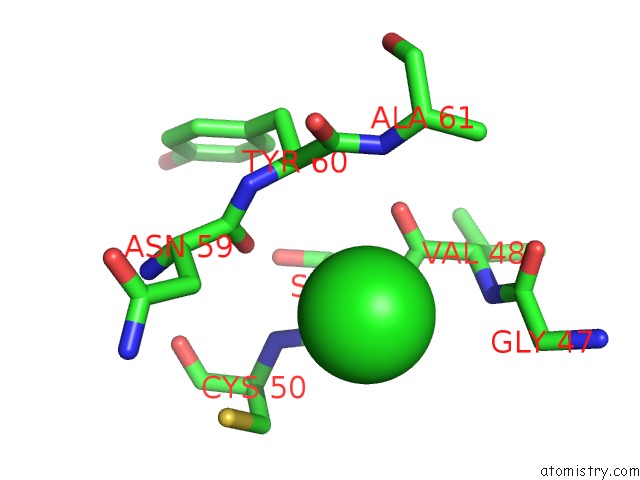
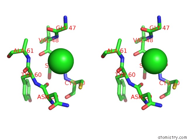
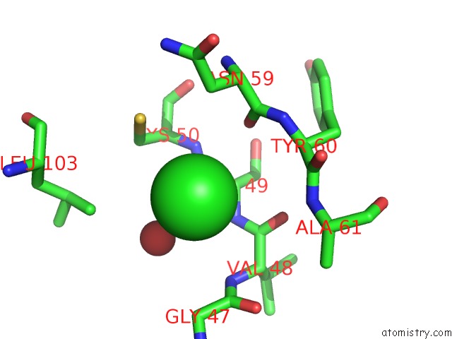
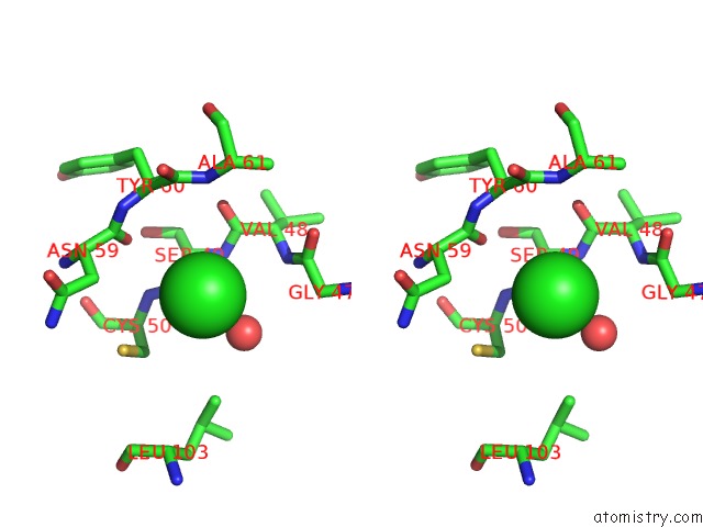
Chlorine binding site 1 out of 4 in 5hdo
Go back to
Chlorine binding site 1 out
of 4 in the Crystal Structure of A Nanobody Raised Against Urokinase-Type Plasminogen Activator
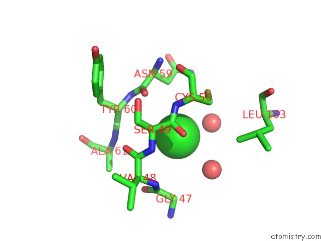
Mono view
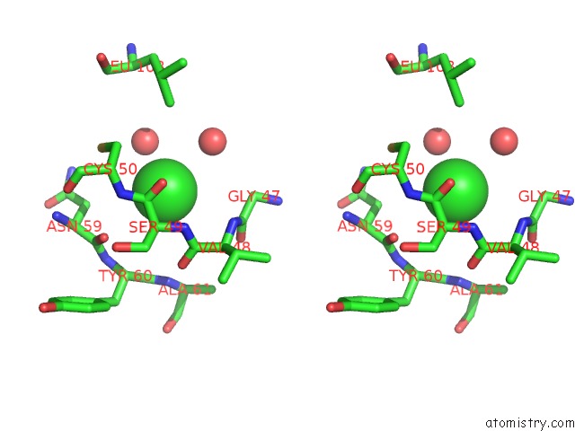
Stereo pair view
Mono view
Stereo pair view
A full contact list of Chlorine with other atoms in the Cl binding
site number 1 of Crystal Structure of A Nanobody Raised Against Urokinase-Type Plasminogen Activator within 5.0Å range:
|
Chlorine binding site 2 out of 4 in 5hdo
Go back to
Chlorine binding site 2 out
of 4 in the Crystal Structure of A Nanobody Raised Against Urokinase-Type Plasminogen Activator
Mono view
Stereo pair view
Mono view
Stereo pair view
A full contact list of Chlorine with other atoms in the Cl binding
site number 2 of Crystal Structure of A Nanobody Raised Against Urokinase-Type Plasminogen Activator within 5.0Å range:
|
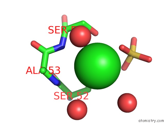
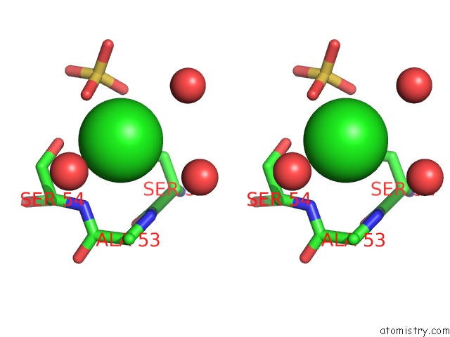
Chlorine binding site 3 out of 4 in 5hdo
Go back to
Chlorine binding site 3 out
of 4 in the Crystal Structure of A Nanobody Raised Against Urokinase-Type Plasminogen Activator
Mono view
Stereo pair view
Mono view
Stereo pair view
A full contact list of Chlorine with other atoms in the Cl binding
site number 3 of Crystal Structure of A Nanobody Raised Against Urokinase-Type Plasminogen Activator within 5.0Å range:
|
Chlorine binding site 4 out of 4 in 5hdo
Go back to
Chlorine binding site 4 out
of 4 in the Crystal Structure of A Nanobody Raised Against Urokinase-Type Plasminogen Activator
Mono view
Stereo pair view
Mono view
Stereo pair view
A full contact list of Chlorine with other atoms in the Cl binding
site number 4 of Crystal Structure of A Nanobody Raised Against Urokinase-Type Plasminogen Activator within 5.0Å range:
|
Reference:
T.Kromann-Hansen,
E.Oldenburg,
K.W.Yung,
G.H.Ghassabeh,
S.Muyldermans,
P.J.Declerck,
M.Huang,
P.A.Andreasen,
J.C.Ngo.
A Camelid-Derived Antibody Fragment Targeting the Active Site of A Serine Protease Balances Between Inhibitor and Substrate Behavior. J.Biol.Chem. V. 291 15156 2016.
ISSN: ESSN 1083-351X
PubMed: 27226628
DOI: 10.1074/JBC.M116.732503
Page generated: Fri Jul 26 08:48:41 2024
ISSN: ESSN 1083-351X
PubMed: 27226628
DOI: 10.1074/JBC.M116.732503
Last articles
Zn in 9J0NZn in 9J0O
Zn in 9J0P
Zn in 9FJX
Zn in 9EKB
Zn in 9C0F
Zn in 9CAH
Zn in 9CH0
Zn in 9CH3
Zn in 9CH1