Chlorine »
PDB 5imx-5ivr »
5iob »
Chlorine in PDB 5iob: Crystal Structure of Beta-N-Acetylglucosaminidase-Like Protein From Corynebacterium Glutamicum
Enzymatic activity of Crystal Structure of Beta-N-Acetylglucosaminidase-Like Protein From Corynebacterium Glutamicum
All present enzymatic activity of Crystal Structure of Beta-N-Acetylglucosaminidase-Like Protein From Corynebacterium Glutamicum:
3.2.1.52;
3.2.1.52;
Protein crystallography data
The structure of Crystal Structure of Beta-N-Acetylglucosaminidase-Like Protein From Corynebacterium Glutamicum, PDB code: 5iob
was solved by
C.Chang,
J.Mack,
M.Endres,
A.Joachimiak,
Midwest Center For Structuralgenomics (Mcsg),
with X-Ray Crystallography technique. A brief refinement statistics is given in the table below:
| Resolution Low / High (Å) | 37.28 / 2.25 |
| Space group | P 21 21 2 |
| Cell size a, b, c (Å), α, β, γ (°) | 308.075, 91.355, 120.018, 90.00, 90.00, 90.00 |
| R / Rfree (%) | 20.9 / 25.7 |
Chlorine Binding Sites:
The binding sites of Chlorine atom in the Crystal Structure of Beta-N-Acetylglucosaminidase-Like Protein From Corynebacterium Glutamicum
(pdb code 5iob). This binding sites where shown within
5.0 Angstroms radius around Chlorine atom.
In total only one binding site of Chlorine was determined in the Crystal Structure of Beta-N-Acetylglucosaminidase-Like Protein From Corynebacterium Glutamicum, PDB code: 5iob:
In total only one binding site of Chlorine was determined in the Crystal Structure of Beta-N-Acetylglucosaminidase-Like Protein From Corynebacterium Glutamicum, PDB code: 5iob:
Chlorine binding site 1 out of 1 in 5iob
Go back to
Chlorine binding site 1 out
of 1 in the Crystal Structure of Beta-N-Acetylglucosaminidase-Like Protein From Corynebacterium Glutamicum
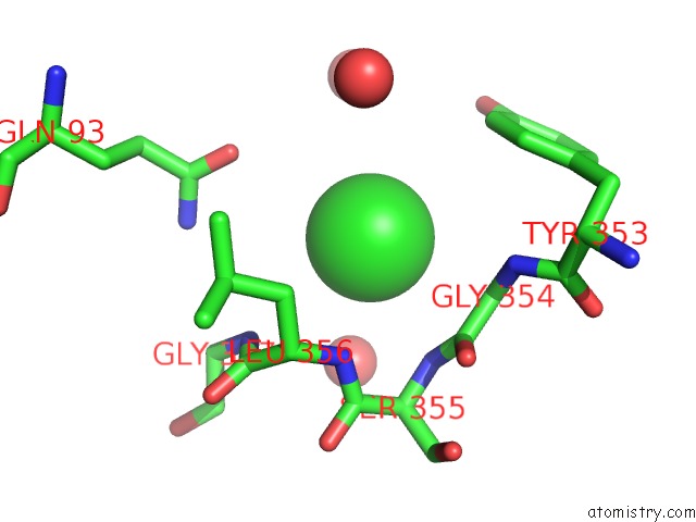
Mono view
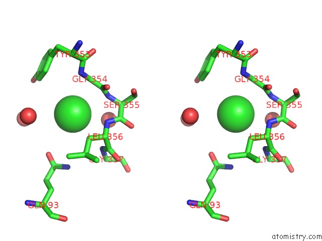
Stereo pair view
Mono view
Stereo pair view
A full contact list of Chlorine with other atoms in the Cl binding
site number 1 of Crystal Structure of Beta-N-Acetylglucosaminidase-Like Protein From Corynebacterium Glutamicum within 5.0Å range:
|
Reference:
C.Chang,
J.Mack,
M.Endres,
A.Joachimiak.
Crystal Structure of Beta-N-Acetylglucosaminidase-Like Protein From Corynebacterium Glutamicum To Be Published.
Page generated: Sat Jul 12 03:14:09 2025
Last articles
Cl in 5RAQCl in 5RAP
Cl in 5RAO
Cl in 5RAN
Cl in 5RAJ
Cl in 5RAM
Cl in 5RAL
Cl in 5RAK
Cl in 5RAF
Cl in 5RAG