Chlorine in PDB 5j57: V5E1-Rta Complex
Enzymatic activity of V5E1-Rta Complex
All present enzymatic activity of V5E1-Rta Complex:
3.2.2.22;
Protein crystallography data
The structure of V5E1-Rta Complex, PDB code: 5j57
was solved by
M.J.Rudolph,
with X-Ray Crystallography technique. A brief refinement statistics is given in the table below:
Resolution Low / High (Å)
|
33.63 /
1.70
|
Space group
|
P 1 21 1
|
Cell size a, b, c (Å), α, β, γ (°)
|
38.692,
68.216,
71.570,
90.00,
92.47,
90.00
|
R / Rfree (%)
|
16.3 /
19
|
Chlorine Binding Sites:
The binding sites of Chlorine atom in the V5E1-Rta Complex
(pdb code 5j57). This binding sites where shown within
5.0 Angstroms radius around Chlorine atom.
In total 5 binding sites of Chlorine where determined in the
V5E1-Rta Complex, PDB code: 5j57:
Jump to Chlorine binding site number:
1;
2;
3;
4;
5;
Chlorine binding site 1 out
of 5 in 5j57
Go back to
Chlorine Binding Sites List in 5j57
Chlorine binding site 1 out
of 5 in the V5E1-Rta Complex
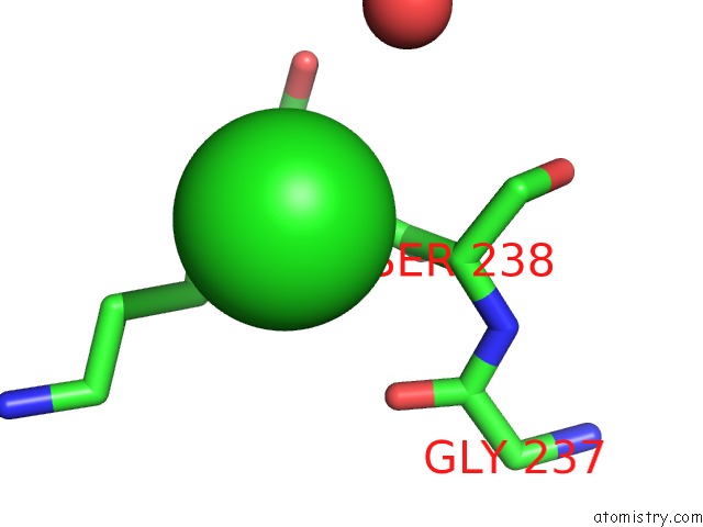
Mono view
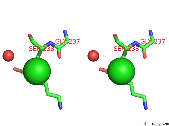
Stereo pair view
|
A full contact list of Chlorine with other atoms in the Cl binding
site number 1 of V5E1-Rta Complex within 5.0Å range:
probe
|
atom
|
residue
|
distance (Å)
|
B
|
Occ
|
A:Cl301
b:33.9
occ:1.00
|
N
|
A:LYS239
|
3.3
|
22.3
|
1.0
|
CA
|
A:SER238
|
3.9
|
21.6
|
1.0
|
CB
|
A:LYS239
|
4.0
|
24.8
|
1.0
|
CG
|
A:LYS239
|
4.0
|
25.8
|
1.0
|
C
|
A:SER238
|
4.1
|
20.9
|
1.0
|
CA
|
A:LYS239
|
4.3
|
22.0
|
1.0
|
CB
|
A:SER238
|
4.4
|
27.7
|
1.0
|
CD
|
A:LYS239
|
4.5
|
33.5
|
1.0
|
O
|
A:HOH494
|
4.6
|
35.7
|
1.0
|
O
|
A:GLY237
|
4.7
|
20.1
|
1.0
|
O
|
A:LYS239
|
4.9
|
26.0
|
1.0
|
N
|
A:SER238
|
5.0
|
20.7
|
1.0
|
|
Chlorine binding site 2 out
of 5 in 5j57
Go back to
Chlorine Binding Sites List in 5j57
Chlorine binding site 2 out
of 5 in the V5E1-Rta Complex
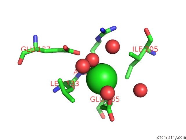
Mono view
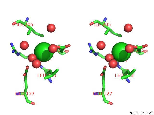
Stereo pair view
|
A full contact list of Chlorine with other atoms in the Cl binding
site number 2 of V5E1-Rta Complex within 5.0Å range:
probe
|
atom
|
residue
|
distance (Å)
|
B
|
Occ
|
A:Cl302
b:35.9
occ:1.00
|
O
|
A:HOH471
|
2.8
|
22.9
|
1.0
|
OE1
|
A:GLU135
|
2.8
|
33.0
|
1.0
|
N
|
A:GLU135
|
2.9
|
16.1
|
1.0
|
CG
|
A:GLU135
|
3.2
|
23.2
|
1.0
|
CB
|
A:GLU135
|
3.4
|
20.2
|
1.0
|
CD1
|
A:ILE205
|
3.4
|
21.5
|
1.0
|
CD
|
A:GLU135
|
3.4
|
30.3
|
1.0
|
N
|
A:ARG134
|
3.5
|
14.7
|
1.0
|
CB
|
A:LEU133
|
3.6
|
15.8
|
1.0
|
CA
|
A:GLU135
|
3.7
|
16.4
|
1.0
|
CB
|
A:ARG134
|
3.8
|
14.3
|
1.0
|
C
|
A:ARG134
|
3.8
|
15.2
|
1.0
|
CA
|
A:ARG134
|
3.9
|
15.3
|
1.0
|
OE2
|
A:GLU127
|
4.0
|
18.2
|
1.0
|
C
|
A:LEU133
|
4.1
|
16.1
|
1.0
|
O
|
A:HOH411
|
4.2
|
45.8
|
1.0
|
CG2
|
A:ILE205
|
4.3
|
18.6
|
1.0
|
CG1
|
A:ILE205
|
4.3
|
18.6
|
1.0
|
CA
|
A:LEU133
|
4.4
|
13.9
|
1.0
|
CD1
|
A:LEU133
|
4.4
|
23.5
|
1.0
|
CG
|
A:LEU133
|
4.5
|
20.5
|
1.0
|
O
|
A:HOH640
|
4.6
|
39.0
|
1.0
|
O
|
A:HOH526
|
4.6
|
21.9
|
1.0
|
O
|
A:HOH602
|
4.6
|
37.5
|
1.0
|
OE2
|
A:GLU135
|
4.7
|
32.1
|
1.0
|
NE
|
A:ARG134
|
4.9
|
14.3
|
1.0
|
CB
|
A:ILE205
|
4.9
|
16.8
|
1.0
|
CD2
|
A:LEU133
|
4.9
|
22.9
|
1.0
|
O
|
A:LEU133
|
4.9
|
15.2
|
1.0
|
O
|
A:ARG134
|
4.9
|
15.3
|
1.0
|
C
|
A:GLU135
|
5.0
|
18.2
|
1.0
|
CD
|
A:GLU127
|
5.0
|
18.6
|
1.0
|
CG
|
A:ARG134
|
5.0
|
14.2
|
1.0
|
|
Chlorine binding site 3 out
of 5 in 5j57
Go back to
Chlorine Binding Sites List in 5j57
Chlorine binding site 3 out
of 5 in the V5E1-Rta Complex
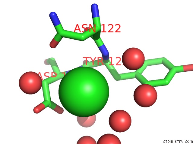
Mono view
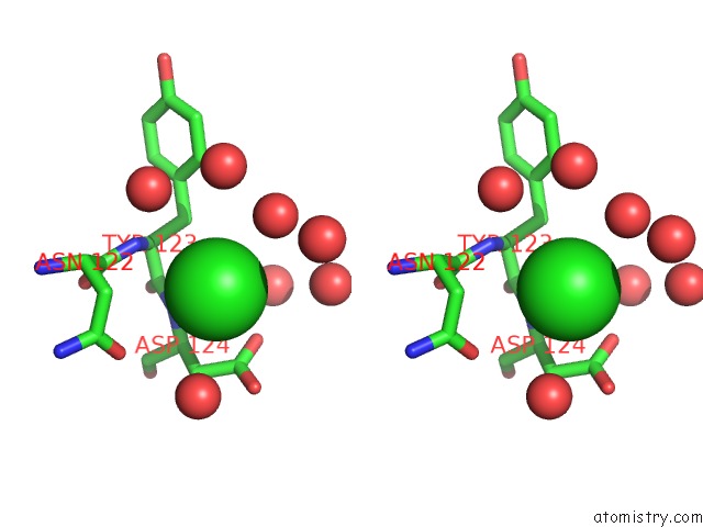
Stereo pair view
|
A full contact list of Chlorine with other atoms in the Cl binding
site number 3 of V5E1-Rta Complex within 5.0Å range:
probe
|
atom
|
residue
|
distance (Å)
|
B
|
Occ
|
A:Cl303
b:45.0
occ:1.00
|
N
|
A:ASP124
|
3.1
|
14.5
|
1.0
|
O
|
A:HOH533
|
3.2
|
32.3
|
1.0
|
CG
|
A:ASP124
|
3.3
|
32.9
|
1.0
|
OD1
|
A:ASP124
|
3.3
|
33.2
|
1.0
|
N
|
A:TYR123
|
3.5
|
15.4
|
1.0
|
CB
|
A:ASP124
|
3.6
|
23.9
|
1.0
|
OD2
|
A:ASP124
|
3.7
|
37.1
|
1.0
|
O
|
A:HOH485
|
3.8
|
48.2
|
1.0
|
CB
|
A:ASN122
|
3.8
|
20.3
|
1.0
|
CA
|
A:ASP124
|
3.9
|
18.7
|
1.0
|
OD1
|
A:ASN122
|
3.9
|
28.8
|
1.0
|
CB
|
A:TYR123
|
3.9
|
13.6
|
1.0
|
CA
|
A:TYR123
|
3.9
|
14.9
|
1.0
|
C
|
A:TYR123
|
3.9
|
13.3
|
1.0
|
O
|
A:HOH557
|
4.1
|
20.0
|
1.0
|
C
|
A:ASN122
|
4.2
|
16.1
|
1.0
|
CG
|
A:ASN122
|
4.3
|
25.5
|
1.0
|
O
|
A:HOH597
|
4.4
|
44.0
|
1.0
|
O
|
A:HOH643
|
4.4
|
22.2
|
1.0
|
CA
|
A:ASN122
|
4.5
|
15.2
|
1.0
|
O
|
A:HOH421
|
4.6
|
23.4
|
1.0
|
O
|
A:HOH588
|
4.8
|
45.0
|
1.0
|
CD2
|
A:TYR123
|
4.9
|
16.6
|
1.0
|
CG
|
A:TYR123
|
4.9
|
14.8
|
1.0
|
O
|
A:ASN122
|
4.9
|
15.2
|
1.0
|
|
Chlorine binding site 4 out
of 5 in 5j57
Go back to
Chlorine Binding Sites List in 5j57
Chlorine binding site 4 out
of 5 in the V5E1-Rta Complex
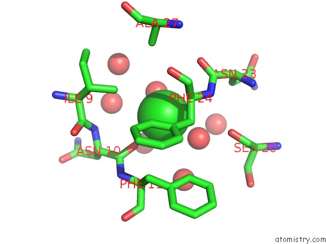
Mono view
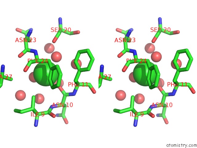
Stereo pair view
|
A full contact list of Chlorine with other atoms in the Cl binding
site number 4 of V5E1-Rta Complex within 5.0Å range:
probe
|
atom
|
residue
|
distance (Å)
|
B
|
Occ
|
A:Cl304
b:37.5
occ:1.00
|
O
|
A:HOH645
|
2.2
|
41.3
|
1.0
|
O
|
A:ASN10
|
2.7
|
20.1
|
1.0
|
O
|
A:HOH474
|
2.9
|
18.3
|
1.0
|
CA
|
A:PHE24
|
3.3
|
10.6
|
1.0
|
CB
|
A:PHE24
|
3.5
|
10.2
|
1.0
|
N
|
A:PHE24
|
3.8
|
12.6
|
1.0
|
CD1
|
A:PHE24
|
3.8
|
14.6
|
1.0
|
C
|
A:ASN10
|
3.9
|
18.6
|
1.0
|
CG2
|
A:ILE9
|
4.0
|
17.2
|
1.0
|
O
|
A:HOH606
|
4.1
|
46.3
|
1.0
|
CG
|
A:PHE24
|
4.1
|
11.3
|
1.0
|
O
|
A:HOH541
|
4.3
|
24.6
|
1.0
|
C
|
A:ASN23
|
4.4
|
13.0
|
1.0
|
O
|
A:ASN23
|
4.5
|
14.6
|
1.0
|
O
|
A:HOH598
|
4.6
|
20.0
|
1.0
|
C
|
A:PHE24
|
4.6
|
12.3
|
1.0
|
N
|
A:ASN10
|
4.6
|
18.6
|
1.0
|
O
|
A:SER20
|
4.6
|
11.7
|
1.0
|
O
|
A:HOH619
|
4.7
|
36.0
|
1.0
|
CA
|
A:PHE11
|
4.8
|
14.3
|
1.0
|
N
|
A:PHE11
|
4.8
|
15.3
|
1.0
|
CB
|
A:PHE11
|
4.8
|
14.1
|
1.0
|
O
|
A:PHE24
|
4.8
|
13.3
|
1.0
|
CD1
|
A:PHE11
|
4.8
|
16.1
|
1.0
|
CA
|
A:ASN10
|
4.9
|
18.5
|
1.0
|
O
|
A:HOH661
|
5.0
|
51.5
|
1.0
|
CB
|
A:ALA27
|
5.0
|
15.6
|
1.0
|
|
Chlorine binding site 5 out
of 5 in 5j57
Go back to
Chlorine Binding Sites List in 5j57
Chlorine binding site 5 out
of 5 in the V5E1-Rta Complex
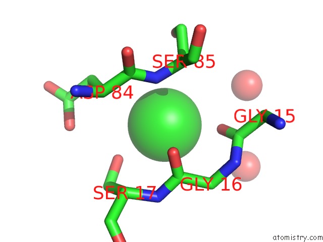
Mono view
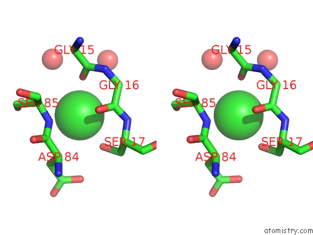
Stereo pair view
|
A full contact list of Chlorine with other atoms in the Cl binding
site number 5 of V5E1-Rta Complex within 5.0Å range:
probe
|
atom
|
residue
|
distance (Å)
|
B
|
Occ
|
B:Cl201
b:47.9
occ:1.00
|
OG
|
B:SER17
|
2.8
|
33.1
|
1.0
|
O
|
B:GLY15
|
2.8
|
30.0
|
1.0
|
N
|
B:SER85
|
3.2
|
28.0
|
1.0
|
O
|
B:GLY16
|
3.6
|
22.7
|
1.0
|
C
|
B:GLY16
|
3.6
|
25.2
|
1.0
|
C
|
B:GLY15
|
3.9
|
28.5
|
1.0
|
CA
|
B:SER85
|
3.9
|
29.4
|
1.0
|
N
|
B:SER17
|
4.0
|
26.2
|
1.0
|
CA
|
B:GLY16
|
4.0
|
26.7
|
1.0
|
CB
|
B:SER17
|
4.0
|
30.8
|
1.0
|
CA
|
B:ASP84
|
4.1
|
28.5
|
1.0
|
C
|
B:ASP84
|
4.1
|
28.7
|
1.0
|
CB
|
B:ASP84
|
4.1
|
30.6
|
1.0
|
OG
|
B:SER85
|
4.4
|
37.0
|
1.0
|
CA
|
B:SER17
|
4.4
|
29.5
|
1.0
|
N
|
B:GLY16
|
4.4
|
25.8
|
1.0
|
OD1
|
B:ASP84
|
4.5
|
33.6
|
1.0
|
O
|
B:HOH371
|
4.5
|
51.5
|
1.0
|
O
|
B:HOH374
|
4.7
|
41.5
|
1.0
|
CG
|
B:ASP84
|
4.7
|
34.0
|
1.0
|
CB
|
B:SER85
|
4.8
|
31.6
|
1.0
|
|
Reference:
M.J.Rudolph,
D.J.Vance,
M.S.Cassidy,
Y.Rong,
N.J.Mantis.
Structural Analysis of Single Domain Antibodies Bound to A Second Neutralizing Hot Spot on Ricin Toxin'S Enzymatic Subunit. J. Biol. Chem. V. 292 872 2017.
ISSN: ESSN 1083-351X
PubMed: 27903650
DOI: 10.1074/JBC.M116.758102
Page generated: Fri Jul 26 09:45:21 2024
|