Chlorine »
PDB 5j7g-5jed »
5j7s »
Chlorine in PDB 5j7s: Crystal Structure of SM1-71 Bound to TAK1-TAB1
Enzymatic activity of Crystal Structure of SM1-71 Bound to TAK1-TAB1
All present enzymatic activity of Crystal Structure of SM1-71 Bound to TAK1-TAB1:
2.7.11.25;
2.7.11.25;
Protein crystallography data
The structure of Crystal Structure of SM1-71 Bound to TAK1-TAB1, PDB code: 5j7s
was solved by
D.Gurbani,
K.D.Westover,
with X-Ray Crystallography technique. A brief refinement statistics is given in the table below:
| Resolution Low / High (Å) | 49.50 / 2.37 |
| Space group | I 2 2 2 |
| Cell size a, b, c (Å), α, β, γ (°) | 58.234, 134.060, 146.814, 90.00, 90.00, 90.00 |
| R / Rfree (%) | 22.8 / 24 |
Chlorine Binding Sites:
The binding sites of Chlorine atom in the Crystal Structure of SM1-71 Bound to TAK1-TAB1
(pdb code 5j7s). This binding sites where shown within
5.0 Angstroms radius around Chlorine atom.
In total only one binding site of Chlorine was determined in the Crystal Structure of SM1-71 Bound to TAK1-TAB1, PDB code: 5j7s:
In total only one binding site of Chlorine was determined in the Crystal Structure of SM1-71 Bound to TAK1-TAB1, PDB code: 5j7s:
Chlorine binding site 1 out of 1 in 5j7s
Go back to
Chlorine binding site 1 out
of 1 in the Crystal Structure of SM1-71 Bound to TAK1-TAB1
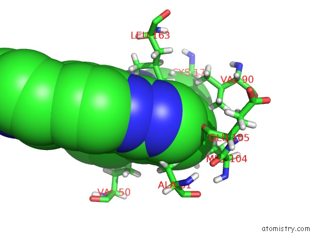
Mono view
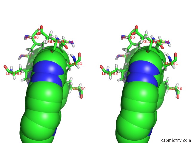
Stereo pair view
Mono view
Stereo pair view
A full contact list of Chlorine with other atoms in the Cl binding
site number 1 of Crystal Structure of SM1-71 Bound to TAK1-TAB1 within 5.0Å range:
|
Reference:
L.Tan,
D.Gurbani,
E.L.Weisberg,
J.C.Hunter,
L.Li,
D.S.Jones,
S.B.Ficarro,
S.Mowafy,
C.P.Tam,
S.Rao,
G.Du,
J.D.Griffin,
P.K.Sorger,
J.A.Marto,
K.D.Westover,
N.S.Gray.
Structure-Guided Development of Covalent TAK1 Inhibitors. Bioorg. Med. Chem. V. 25 838 2017.
ISSN: ESSN 1464-3391
PubMed: 28011204
DOI: 10.1016/J.BMC.2016.11.035
Page generated: Fri Jul 26 09:49:45 2024
ISSN: ESSN 1464-3391
PubMed: 28011204
DOI: 10.1016/J.BMC.2016.11.035
Last articles
Zn in 9J0NZn in 9J0O
Zn in 9J0P
Zn in 9FJX
Zn in 9EKB
Zn in 9C0F
Zn in 9CAH
Zn in 9CH0
Zn in 9CH3
Zn in 9CH1