Chlorine »
PDB 5jen-5jkc »
5jhb »
Chlorine in PDB 5jhb: Structure of Phosphoinositide 3-Kinase Gamma (PI3K) Bound to the Potent Inhibitor PIKIN3
Enzymatic activity of Structure of Phosphoinositide 3-Kinase Gamma (PI3K) Bound to the Potent Inhibitor PIKIN3
All present enzymatic activity of Structure of Phosphoinositide 3-Kinase Gamma (PI3K) Bound to the Potent Inhibitor PIKIN3:
2.7.1.153; 2.7.11.1;
2.7.1.153; 2.7.11.1;
Protein crystallography data
The structure of Structure of Phosphoinositide 3-Kinase Gamma (PI3K) Bound to the Potent Inhibitor PIKIN3, PDB code: 5jhb
was solved by
J.E.Burke,
A.J.Inglis,
R.L.Williams,
with X-Ray Crystallography technique. A brief refinement statistics is given in the table below:
| Resolution Low / High (Å) | 62.08 / 2.48 |
| Space group | C 1 2 1 |
| Cell size a, b, c (Å), α, β, γ (°) | 142.530, 67.600, 106.550, 90.00, 96.23, 90.00 |
| R / Rfree (%) | 23.1 / 27.7 |
Other elements in 5jhb:
The structure of Structure of Phosphoinositide 3-Kinase Gamma (PI3K) Bound to the Potent Inhibitor PIKIN3 also contains other interesting chemical elements:
| Fluorine | (F) | 3 atoms |
Chlorine Binding Sites:
The binding sites of Chlorine atom in the Structure of Phosphoinositide 3-Kinase Gamma (PI3K) Bound to the Potent Inhibitor PIKIN3
(pdb code 5jhb). This binding sites where shown within
5.0 Angstroms radius around Chlorine atom.
In total only one binding site of Chlorine was determined in the Structure of Phosphoinositide 3-Kinase Gamma (PI3K) Bound to the Potent Inhibitor PIKIN3, PDB code: 5jhb:
In total only one binding site of Chlorine was determined in the Structure of Phosphoinositide 3-Kinase Gamma (PI3K) Bound to the Potent Inhibitor PIKIN3, PDB code: 5jhb:
Chlorine binding site 1 out of 1 in 5jhb
Go back to
Chlorine binding site 1 out
of 1 in the Structure of Phosphoinositide 3-Kinase Gamma (PI3K) Bound to the Potent Inhibitor PIKIN3
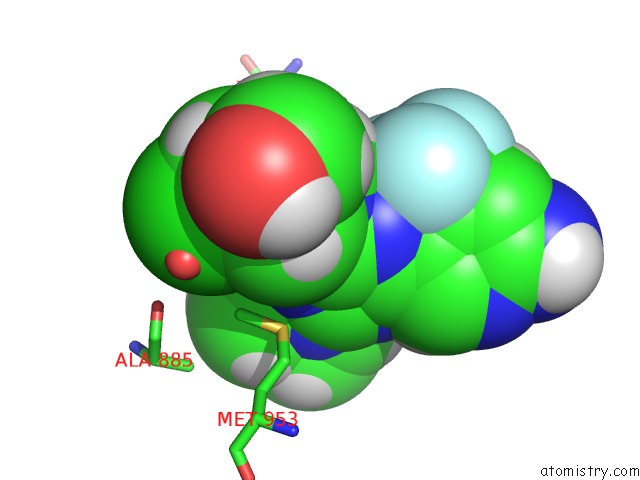
Mono view
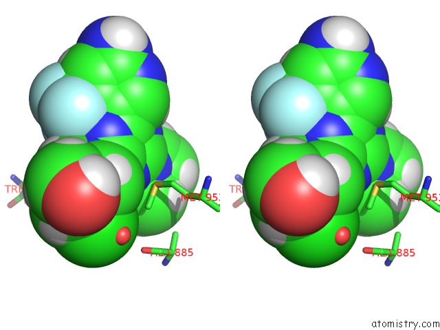
Stereo pair view
Mono view
Stereo pair view
A full contact list of Chlorine with other atoms in the Cl binding
site number 1 of Structure of Phosphoinositide 3-Kinase Gamma (PI3K) Bound to the Potent Inhibitor PIKIN3 within 5.0Å range:
|
Reference:
T.Bohnacker,
A.E.Prota,
F.Beaufils,
J.E.Burke,
A.Melone,
A.J.Inglis,
D.Rageot,
A.M.Sele,
V.Cmiljanovic,
N.Cmiljanovic,
K.Bargsten,
A.Aher,
A.Akhmanova,
J.F.Diaz,
D.Fabbro,
M.Zvelebil,
R.L.Williams,
M.O.Steinmetz,
M.P.Wymann.
Deconvolution of Buparlisib'S Mechanism of Action Defines Specific PI3K and Tubulin Inhibitors For Therapeutic Intervention. Nat Commun V. 8 14683 2017.
ISSN: ESSN 2041-1723
PubMed: 28276440
DOI: 10.1038/NCOMMS14683
Page generated: Sat Jul 12 03:38:08 2025
ISSN: ESSN 2041-1723
PubMed: 28276440
DOI: 10.1038/NCOMMS14683
Last articles
Fe in 2YXOFe in 2YRS
Fe in 2YXC
Fe in 2YNM
Fe in 2YVJ
Fe in 2YP1
Fe in 2YU2
Fe in 2YU1
Fe in 2YQB
Fe in 2YOO