Chlorine »
PDB 5jen-5jkc »
5jk0 »
Chlorine in PDB 5jk0: Crystal Structure of Xerh Site-Specific Recombinase Bound to Difh Substrate: Pre-Cleavage Complex
Protein crystallography data
The structure of Crystal Structure of Xerh Site-Specific Recombinase Bound to Difh Substrate: Pre-Cleavage Complex, PDB code: 5jk0
was solved by
A.Bebel,
O.Barabas,
with X-Ray Crystallography technique. A brief refinement statistics is given in the table below:
| Resolution Low / High (Å) | 48.89 / 2.10 |
| Space group | P 21 21 21 |
| Cell size a, b, c (Å), α, β, γ (°) | 79.280, 153.200, 169.390, 90.00, 90.00, 90.00 |
| R / Rfree (%) | 19.1 / 22.3 |
Chlorine Binding Sites:
The binding sites of Chlorine atom in the Crystal Structure of Xerh Site-Specific Recombinase Bound to Difh Substrate: Pre-Cleavage Complex
(pdb code 5jk0). This binding sites where shown within
5.0 Angstroms radius around Chlorine atom.
In total only one binding site of Chlorine was determined in the Crystal Structure of Xerh Site-Specific Recombinase Bound to Difh Substrate: Pre-Cleavage Complex, PDB code: 5jk0:
In total only one binding site of Chlorine was determined in the Crystal Structure of Xerh Site-Specific Recombinase Bound to Difh Substrate: Pre-Cleavage Complex, PDB code: 5jk0:
Chlorine binding site 1 out of 1 in 5jk0
Go back to
Chlorine binding site 1 out
of 1 in the Crystal Structure of Xerh Site-Specific Recombinase Bound to Difh Substrate: Pre-Cleavage Complex
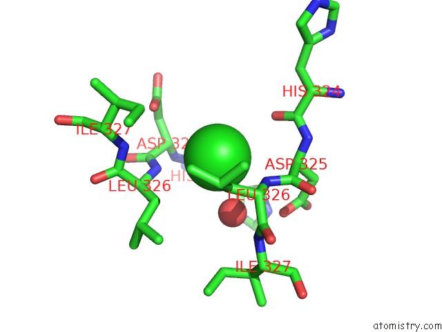
Mono view
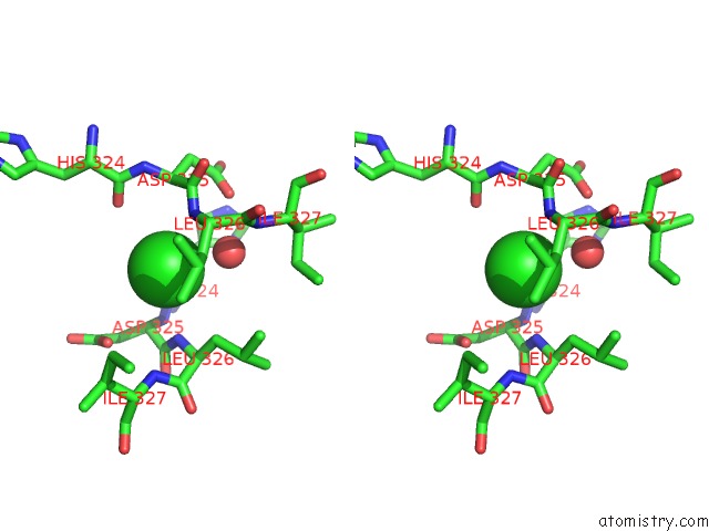
Stereo pair view
Mono view
Stereo pair view
A full contact list of Chlorine with other atoms in the Cl binding
site number 1 of Crystal Structure of Xerh Site-Specific Recombinase Bound to Difh Substrate: Pre-Cleavage Complex within 5.0Å range:
|
Reference:
A.Bebel,
E.Karaca,
B.Kumar,
W.M.Stark,
O.Barabas.
Structural Snapshots of Xer Recombination Reveal Activation By Synaptic Complex Remodeling and Dna Bending. Elife V. 5 2016.
ISSN: ESSN 2050-084X
PubMed: 28009253
DOI: 10.7554/ELIFE.19706
Page generated: Fri Jul 26 10:06:02 2024
ISSN: ESSN 2050-084X
PubMed: 28009253
DOI: 10.7554/ELIFE.19706
Last articles
Zn in 9J0NZn in 9J0O
Zn in 9J0P
Zn in 9FJX
Zn in 9EKB
Zn in 9C0F
Zn in 9CAH
Zn in 9CH0
Zn in 9CH3
Zn in 9CH1