Chlorine »
PDB 5jkd-5jsv »
5jmr »
Chlorine in PDB 5jmr: X-Ray Structure of the Furin Inhibitory Antibody NB14
Protein crystallography data
The structure of X-Ray Structure of the Furin Inhibitory Antibody NB14, PDB code: 5jmr
was solved by
S.O.Dahms,
M.E.Than,
with X-Ray Crystallography technique. A brief refinement statistics is given in the table below:
| Resolution Low / High (Å) | 35.36 / 2.27 |
| Space group | I 41 2 2 |
| Cell size a, b, c (Å), α, β, γ (°) | 91.411, 91.411, 211.290, 90.00, 90.00, 90.00 |
| R / Rfree (%) | 19 / 21.6 |
Chlorine Binding Sites:
The binding sites of Chlorine atom in the X-Ray Structure of the Furin Inhibitory Antibody NB14
(pdb code 5jmr). This binding sites where shown within
5.0 Angstroms radius around Chlorine atom.
In total 2 binding sites of Chlorine where determined in the X-Ray Structure of the Furin Inhibitory Antibody NB14, PDB code: 5jmr:
Jump to Chlorine binding site number: 1; 2;
In total 2 binding sites of Chlorine where determined in the X-Ray Structure of the Furin Inhibitory Antibody NB14, PDB code: 5jmr:
Jump to Chlorine binding site number: 1; 2;
Chlorine binding site 1 out of 2 in 5jmr
Go back to
Chlorine binding site 1 out
of 2 in the X-Ray Structure of the Furin Inhibitory Antibody NB14

Mono view
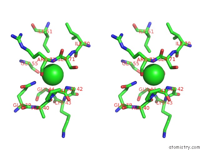
Stereo pair view
Mono view
Stereo pair view
A full contact list of Chlorine with other atoms in the Cl binding
site number 1 of X-Ray Structure of the Furin Inhibitory Antibody NB14 within 5.0Å range:
|
Chlorine binding site 2 out of 2 in 5jmr
Go back to
Chlorine binding site 2 out
of 2 in the X-Ray Structure of the Furin Inhibitory Antibody NB14
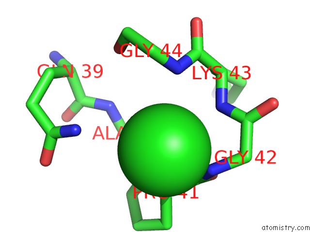
Mono view
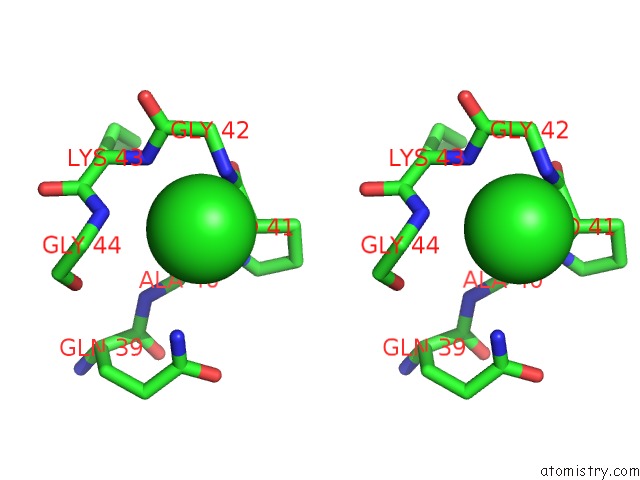
Stereo pair view
Mono view
Stereo pair view
A full contact list of Chlorine with other atoms in the Cl binding
site number 2 of X-Ray Structure of the Furin Inhibitory Antibody NB14 within 5.0Å range:
|
Reference:
S.O.Dahms,
J.W.Creemers,
Y.Schaub,
G.P.Bourenkov,
T.Zogg,
H.Brandstetter,
M.E.Than.
The Structure of A Furin-Antibody Complex Explains Non-Competitive Inhibition By Steric Exclusion of Substrate Conformers. Sci Rep V. 6 34303 2016.
ISSN: ESSN 2045-2322
PubMed: 27670069
DOI: 10.1038/SREP34303
Page generated: Fri Jul 26 10:08:54 2024
ISSN: ESSN 2045-2322
PubMed: 27670069
DOI: 10.1038/SREP34303
Last articles
Zn in 9J0NZn in 9J0O
Zn in 9J0P
Zn in 9FJX
Zn in 9EKB
Zn in 9C0F
Zn in 9CAH
Zn in 9CH0
Zn in 9CH3
Zn in 9CH1