Chlorine »
PDB 5jsw-5jzn »
5jxf »
Chlorine in PDB 5jxf: Crystal Structure of Flavobacterium Psychrophilum DPP11 in Complex with Dipeptide Arg-Asp
Protein crystallography data
The structure of Crystal Structure of Flavobacterium Psychrophilum DPP11 in Complex with Dipeptide Arg-Asp, PDB code: 5jxf
was solved by
G.A.Bezerra,
S.Fedosyuk,
Y.Ohara-Nemoto,
T.K.Nemoto,
K.Djinovic-Carugo,
with X-Ray Crystallography technique. A brief refinement statistics is given in the table below:
| Resolution Low / High (Å) | 46.83 / 2.10 |
| Space group | P 1 21 1 |
| Cell size a, b, c (Å), α, β, γ (°) | 126.051, 70.689, 191.598, 90.00, 97.26, 90.00 |
| R / Rfree (%) | 19.6 / 24.4 |
Other elements in 5jxf:
The structure of Crystal Structure of Flavobacterium Psychrophilum DPP11 in Complex with Dipeptide Arg-Asp also contains other interesting chemical elements:
| Sodium | (Na) | 1 atom |
Chlorine Binding Sites:
The binding sites of Chlorine atom in the Crystal Structure of Flavobacterium Psychrophilum DPP11 in Complex with Dipeptide Arg-Asp
(pdb code 5jxf). This binding sites where shown within
5.0 Angstroms radius around Chlorine atom.
In total 3 binding sites of Chlorine where determined in the Crystal Structure of Flavobacterium Psychrophilum DPP11 in Complex with Dipeptide Arg-Asp, PDB code: 5jxf:
Jump to Chlorine binding site number: 1; 2; 3;
In total 3 binding sites of Chlorine where determined in the Crystal Structure of Flavobacterium Psychrophilum DPP11 in Complex with Dipeptide Arg-Asp, PDB code: 5jxf:
Jump to Chlorine binding site number: 1; 2; 3;
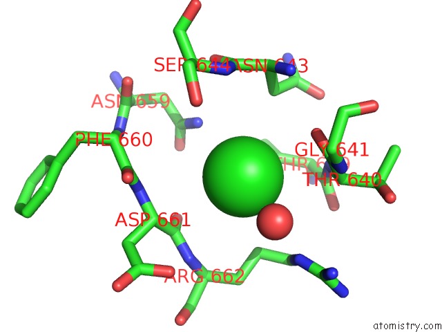
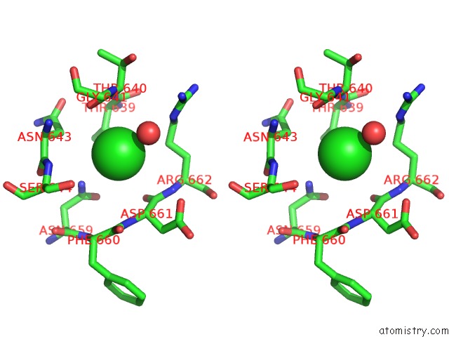
Chlorine binding site 1 out of 3 in 5jxf
Go back to
Chlorine binding site 1 out
of 3 in the Crystal Structure of Flavobacterium Psychrophilum DPP11 in Complex with Dipeptide Arg-Asp
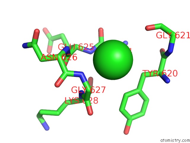
Mono view
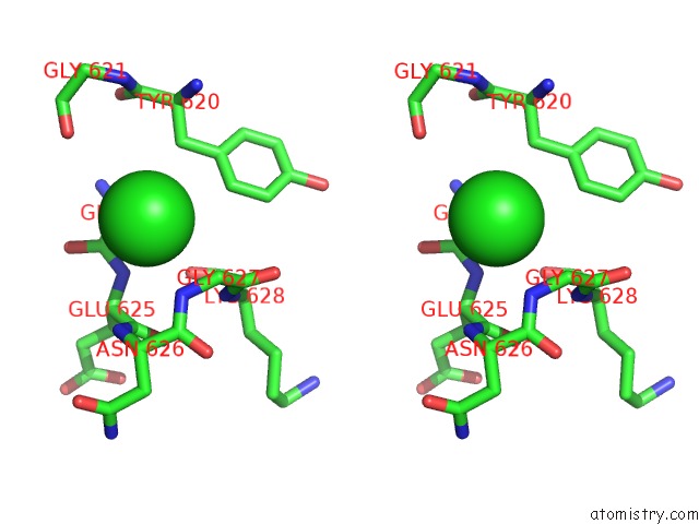
Stereo pair view
Mono view
Stereo pair view
A full contact list of Chlorine with other atoms in the Cl binding
site number 1 of Crystal Structure of Flavobacterium Psychrophilum DPP11 in Complex with Dipeptide Arg-Asp within 5.0Å range:
|
Chlorine binding site 2 out of 3 in 5jxf
Go back to
Chlorine binding site 2 out
of 3 in the Crystal Structure of Flavobacterium Psychrophilum DPP11 in Complex with Dipeptide Arg-Asp
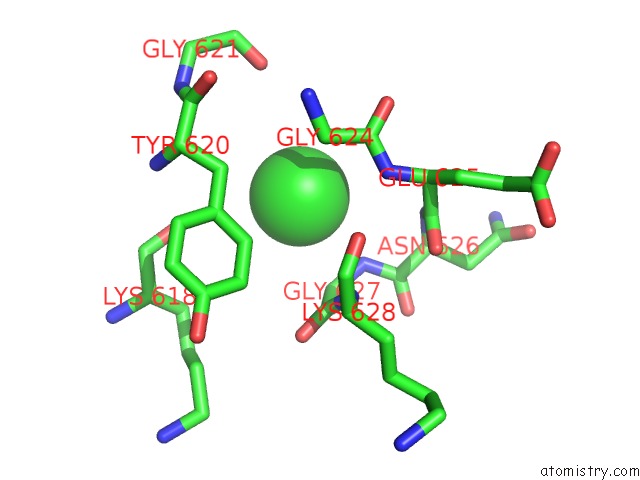
Mono view
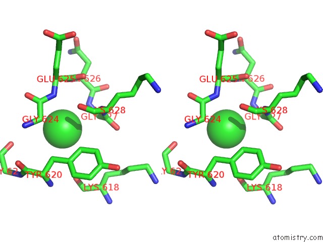
Stereo pair view
Mono view
Stereo pair view
A full contact list of Chlorine with other atoms in the Cl binding
site number 2 of Crystal Structure of Flavobacterium Psychrophilum DPP11 in Complex with Dipeptide Arg-Asp within 5.0Å range:
|
Chlorine binding site 3 out of 3 in 5jxf
Go back to
Chlorine binding site 3 out
of 3 in the Crystal Structure of Flavobacterium Psychrophilum DPP11 in Complex with Dipeptide Arg-Asp
Mono view
Stereo pair view
Mono view
Stereo pair view
A full contact list of Chlorine with other atoms in the Cl binding
site number 3 of Crystal Structure of Flavobacterium Psychrophilum DPP11 in Complex with Dipeptide Arg-Asp within 5.0Å range:
|
Reference:
G.A.Bezerra,
Y.Ohara-Nemoto,
I.Cornaciu,
S.Fedosyuk,
G.Hoffmann,
A.Round,
J.A.Marquez,
T.K.Nemoto,
K.Djinovic-Carugo.
Bacterial Protease Uses Distinct Thermodynamic Signatures For Substrate Recognition. Sci Rep V. 7 2848 2017.
ISSN: ESSN 2045-2322
PubMed: 28588213
DOI: 10.1038/S41598-017-03220-Y
Page generated: Sat Jul 12 03:47:31 2025
ISSN: ESSN 2045-2322
PubMed: 28588213
DOI: 10.1038/S41598-017-03220-Y
Last articles
Fe in 2YXOFe in 2YRS
Fe in 2YXC
Fe in 2YNM
Fe in 2YVJ
Fe in 2YP1
Fe in 2YU2
Fe in 2YU1
Fe in 2YQB
Fe in 2YOO