Chlorine »
PDB 5jsw-5jzn »
5jxk »
Chlorine in PDB 5jxk: Crystal Structure of Porphyromonas Endodontalis DPP11
Protein crystallography data
The structure of Crystal Structure of Porphyromonas Endodontalis DPP11, PDB code: 5jxk
was solved by
G.A.Bezerra,
I.Cornaciu,
G.Hoffmann,
K.Djinovic-Carugo,
J.A.Marquez,
with X-Ray Crystallography technique. A brief refinement statistics is given in the table below:
| Resolution Low / High (Å) | 46.70 / 2.85 |
| Space group | P 21 21 21 |
| Cell size a, b, c (Å), α, β, γ (°) | 76.752, 91.831, 229.917, 90.00, 90.00, 90.00 |
| R / Rfree (%) | 24 / 27.3 |
Chlorine Binding Sites:
The binding sites of Chlorine atom in the Crystal Structure of Porphyromonas Endodontalis DPP11
(pdb code 5jxk). This binding sites where shown within
5.0 Angstroms radius around Chlorine atom.
In total 3 binding sites of Chlorine where determined in the Crystal Structure of Porphyromonas Endodontalis DPP11, PDB code: 5jxk:
Jump to Chlorine binding site number: 1; 2; 3;
In total 3 binding sites of Chlorine where determined in the Crystal Structure of Porphyromonas Endodontalis DPP11, PDB code: 5jxk:
Jump to Chlorine binding site number: 1; 2; 3;
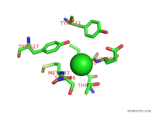
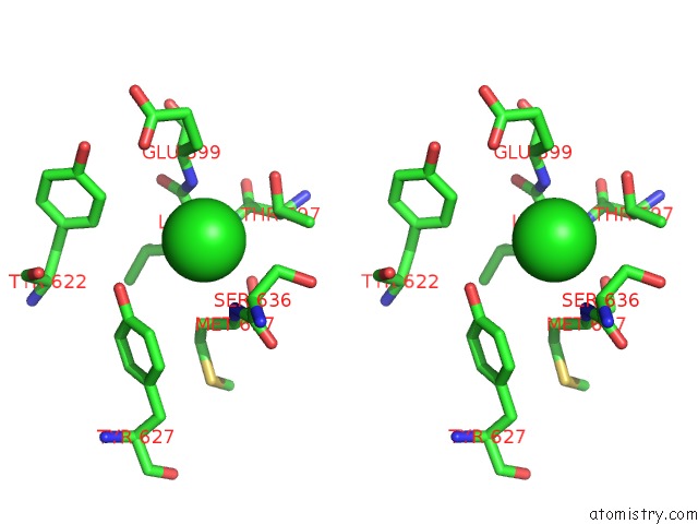
Chlorine binding site 1 out of 3 in 5jxk
Go back to
Chlorine binding site 1 out
of 3 in the Crystal Structure of Porphyromonas Endodontalis DPP11
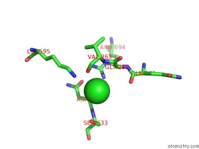
Mono view
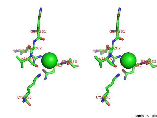
Stereo pair view
Mono view
Stereo pair view
A full contact list of Chlorine with other atoms in the Cl binding
site number 1 of Crystal Structure of Porphyromonas Endodontalis DPP11 within 5.0Å range:
|
Chlorine binding site 2 out of 3 in 5jxk
Go back to
Chlorine binding site 2 out
of 3 in the Crystal Structure of Porphyromonas Endodontalis DPP11
Mono view
Stereo pair view
Mono view
Stereo pair view
A full contact list of Chlorine with other atoms in the Cl binding
site number 2 of Crystal Structure of Porphyromonas Endodontalis DPP11 within 5.0Å range:
|
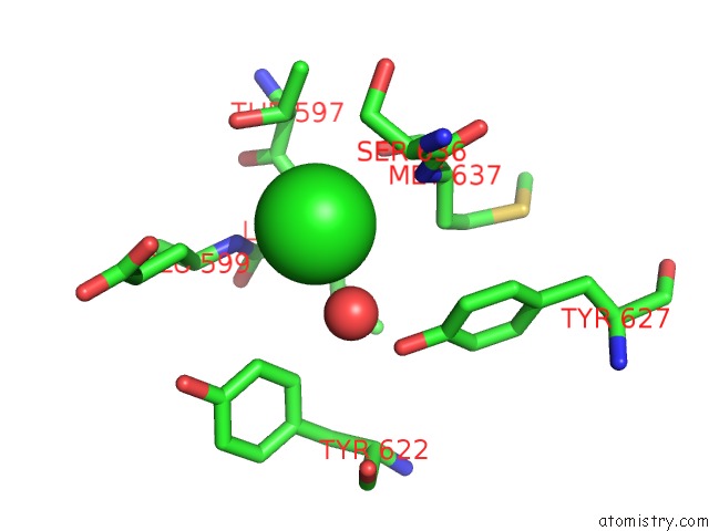
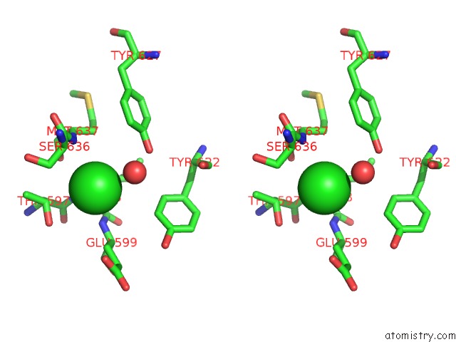
Chlorine binding site 3 out of 3 in 5jxk
Go back to
Chlorine binding site 3 out
of 3 in the Crystal Structure of Porphyromonas Endodontalis DPP11
Mono view
Stereo pair view
Mono view
Stereo pair view
A full contact list of Chlorine with other atoms in the Cl binding
site number 3 of Crystal Structure of Porphyromonas Endodontalis DPP11 within 5.0Å range:
|
Reference:
G.A.Bezerra,
Y.Ohara-Nemoto,
I.Cornaciu,
S.Fedosyuk,
G.Hoffmann,
A.Round,
J.A.Marquez,
T.K.Nemoto,
K.Djinovic-Carugo.
Bacterial Protease Uses Distinct Thermodynamic Signatures For Substrate Recognition. Sci Rep V. 7 2848 2017.
ISSN: ESSN 2045-2322
PubMed: 28588213
DOI: 10.1038/S41598-017-03220-Y
Page generated: Fri Jul 26 10:18:06 2024
ISSN: ESSN 2045-2322
PubMed: 28588213
DOI: 10.1038/S41598-017-03220-Y
Last articles
Zn in 9J0NZn in 9J0O
Zn in 9J0P
Zn in 9FJX
Zn in 9EKB
Zn in 9C0F
Zn in 9CAH
Zn in 9CH0
Zn in 9CH3
Zn in 9CH1