Chlorine »
PDB 5jsw-5jzn »
5jxr »
Chlorine in PDB 5jxr: Crystal Structure of Mtiswi
Protein crystallography data
The structure of Crystal Structure of Mtiswi, PDB code: 5jxr
was solved by
Z.Chen,
L.Yan,
with X-Ray Crystallography technique. A brief refinement statistics is given in the table below:
| Resolution Low / High (Å) | 32.91 / 2.40 |
| Space group | P 31 |
| Cell size a, b, c (Å), α, β, γ (°) | 127.767, 127.767, 106.666, 90.00, 90.00, 120.00 |
| R / Rfree (%) | 19.5 / 22.6 |
Chlorine Binding Sites:
The binding sites of Chlorine atom in the Crystal Structure of Mtiswi
(pdb code 5jxr). This binding sites where shown within
5.0 Angstroms radius around Chlorine atom.
In total 2 binding sites of Chlorine where determined in the Crystal Structure of Mtiswi, PDB code: 5jxr:
Jump to Chlorine binding site number: 1; 2;
In total 2 binding sites of Chlorine where determined in the Crystal Structure of Mtiswi, PDB code: 5jxr:
Jump to Chlorine binding site number: 1; 2;
Chlorine binding site 1 out of 2 in 5jxr
Go back to
Chlorine binding site 1 out
of 2 in the Crystal Structure of Mtiswi
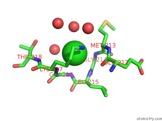
Mono view
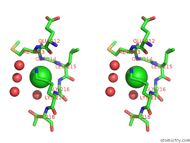
Stereo pair view
Mono view
Stereo pair view
A full contact list of Chlorine with other atoms in the Cl binding
site number 1 of Crystal Structure of Mtiswi within 5.0Å range:
|
Chlorine binding site 2 out of 2 in 5jxr
Go back to
Chlorine binding site 2 out
of 2 in the Crystal Structure of Mtiswi
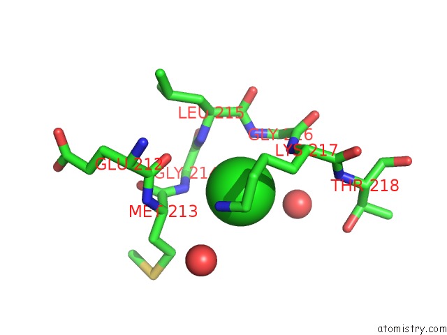
Mono view
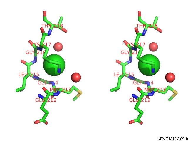
Stereo pair view
Mono view
Stereo pair view
A full contact list of Chlorine with other atoms in the Cl binding
site number 2 of Crystal Structure of Mtiswi within 5.0Å range:
|
Reference:
L.Yan,
L.Wang,
Y.Tian,
X.Xia,
Z.Chen.
Structure and Regulation of the Chromatin Remodeller Iswi Nature V. 540 466 2016.
ISSN: ESSN 1476-4687
PubMed: 27919072
DOI: 10.1038/NATURE20590
Page generated: Fri Jul 26 10:18:20 2024
ISSN: ESSN 1476-4687
PubMed: 27919072
DOI: 10.1038/NATURE20590
Last articles
Zn in 9J0NZn in 9J0O
Zn in 9J0P
Zn in 9FJX
Zn in 9EKB
Zn in 9C0F
Zn in 9CAH
Zn in 9CH0
Zn in 9CH3
Zn in 9CH1