Chlorine »
PDB 5kgg-5kp3 »
5klv »
Chlorine in PDB 5klv: Structure of Bos Taurus Cytochrome BC1 with Fenamidone Inhibited
Enzymatic activity of Structure of Bos Taurus Cytochrome BC1 with Fenamidone Inhibited
All present enzymatic activity of Structure of Bos Taurus Cytochrome BC1 with Fenamidone Inhibited:
1.10.2.2;
1.10.2.2;
Protein crystallography data
The structure of Structure of Bos Taurus Cytochrome BC1 with Fenamidone Inhibited, PDB code: 5klv
was solved by
D.Xia,
L.Esser,
F.Zhou,
Y.Zhou,
Y.Xiao,
W.K.Tang,
C.A.Yu,
Z.Qin,
with X-Ray Crystallography technique. A brief refinement statistics is given in the table below:
| Resolution Low / High (Å) | 34.68 / 2.65 |
| Space group | I 41 2 2 |
| Cell size a, b, c (Å), α, β, γ (°) | 153.986, 153.986, 592.713, 90.00, 90.00, 90.00 |
| R / Rfree (%) | 22.8 / 26.9 |
Other elements in 5klv:
The structure of Structure of Bos Taurus Cytochrome BC1 with Fenamidone Inhibited also contains other interesting chemical elements:
| Iron | (Fe) | 5 atoms |
Chlorine Binding Sites:
The binding sites of Chlorine atom in the Structure of Bos Taurus Cytochrome BC1 with Fenamidone Inhibited
(pdb code 5klv). This binding sites where shown within
5.0 Angstroms radius around Chlorine atom.
In total only one binding site of Chlorine was determined in the Structure of Bos Taurus Cytochrome BC1 with Fenamidone Inhibited, PDB code: 5klv:
In total only one binding site of Chlorine was determined in the Structure of Bos Taurus Cytochrome BC1 with Fenamidone Inhibited, PDB code: 5klv:
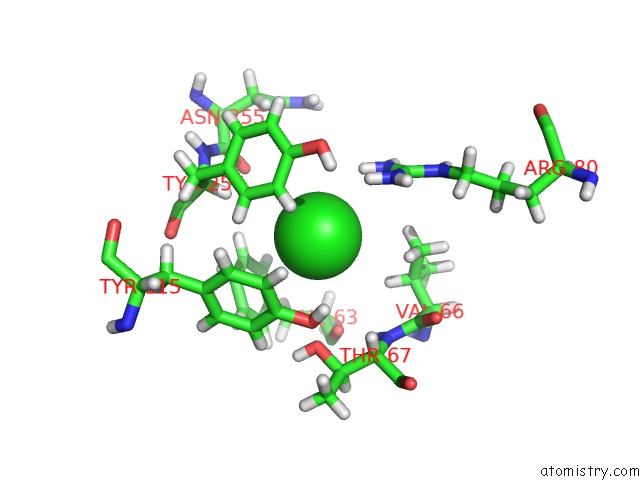
Chlorine binding site 1 out of 1 in 5klv
Go back to
Chlorine binding site 1 out
of 1 in the Structure of Bos Taurus Cytochrome BC1 with Fenamidone Inhibited
Mono view

Stereo pair view
Mono view
Stereo pair view
A full contact list of Chlorine with other atoms in the Cl binding
site number 1 of Structure of Bos Taurus Cytochrome BC1 with Fenamidone Inhibited within 5.0Å range:
|
Reference:
L.Esser,
F.Zhou,
Y.Zhou,
Y.Xiao,
W.K.Tang,
C.A.Yu,
Z.Qin,
D.Xia.
Hydrogen Bonding to the Substrate Is Not Required For Rieske Iron-Sulfur Protein Docking to the Quinol Oxidation Site of Complex III. J.Biol.Chem. V. 291 25019 2016.
ISSN: ESSN 1083-351X
PubMed: 27758861
DOI: 10.1074/JBC.M116.744391
Page generated: Sat Jul 12 04:06:31 2025
ISSN: ESSN 1083-351X
PubMed: 27758861
DOI: 10.1074/JBC.M116.744391
Last articles
Fe in 2YXOFe in 2YRS
Fe in 2YXC
Fe in 2YNM
Fe in 2YVJ
Fe in 2YP1
Fe in 2YU2
Fe in 2YU1
Fe in 2YQB
Fe in 2YOO