Chlorine »
PDB 5kq0-5kvy »
5ku1 »
Chlorine in PDB 5ku1: HMIRO1 Ef Hand and Cgtpase Domains in the Gdp-Bound State
Protein crystallography data
The structure of HMIRO1 Ef Hand and Cgtpase Domains in the Gdp-Bound State, PDB code: 5ku1
was solved by
J.L.Klosowiak,
P.J.Focia,
S.E.Rice,
D.M.Freymann,
with X-Ray Crystallography technique. A brief refinement statistics is given in the table below:
| Resolution Low / High (Å) | 29.81 / 2.50 |
| Space group | P 43 21 2 |
| Cell size a, b, c (Å), α, β, γ (°) | 73.136, 73.136, 217.142, 90.00, 90.00, 90.00 |
| R / Rfree (%) | 22.6 / 26.4 |
Other elements in 5ku1:
The structure of HMIRO1 Ef Hand and Cgtpase Domains in the Gdp-Bound State also contains other interesting chemical elements:
| Magnesium | (Mg) | 2 atoms |
Chlorine Binding Sites:
The binding sites of Chlorine atom in the HMIRO1 Ef Hand and Cgtpase Domains in the Gdp-Bound State
(pdb code 5ku1). This binding sites where shown within
5.0 Angstroms radius around Chlorine atom.
In total only one binding site of Chlorine was determined in the HMIRO1 Ef Hand and Cgtpase Domains in the Gdp-Bound State, PDB code: 5ku1:
In total only one binding site of Chlorine was determined in the HMIRO1 Ef Hand and Cgtpase Domains in the Gdp-Bound State, PDB code: 5ku1:
Chlorine binding site 1 out of 1 in 5ku1
Go back to
Chlorine binding site 1 out
of 1 in the HMIRO1 Ef Hand and Cgtpase Domains in the Gdp-Bound State
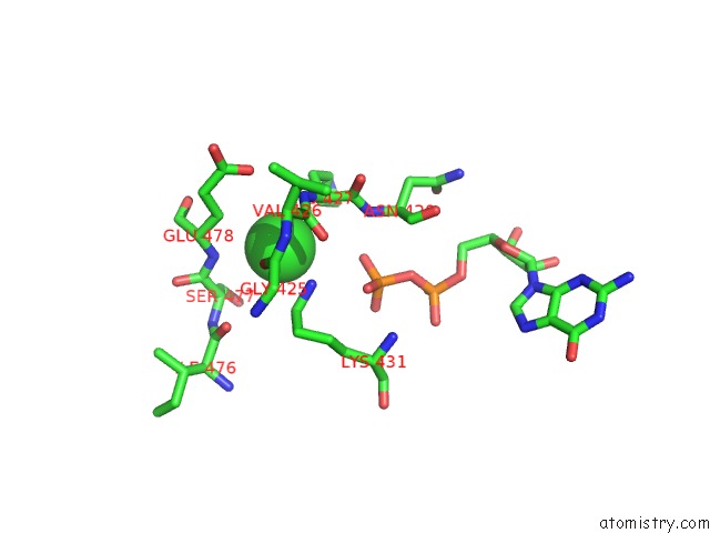
Mono view
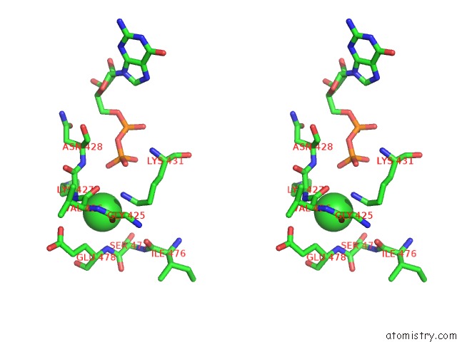
Stereo pair view
Mono view
Stereo pair view
A full contact list of Chlorine with other atoms in the Cl binding
site number 1 of HMIRO1 Ef Hand and Cgtpase Domains in the Gdp-Bound State within 5.0Å range:
|
Reference:
J.L.Klosowiak,
S.Park,
K.P.Smith,
M.E.French,
P.J.Focia,
D.M.Freymann,
S.E.Rice.
Structural Insights Into Parkin Substrate Lysine Targeting From Minimal Miro Substrates. Sci Rep V. 6 33019 2016.
ISSN: ESSN 2045-2322
PubMed: 27605430
DOI: 10.1038/SREP33019
Page generated: Fri Jul 26 10:54:17 2024
ISSN: ESSN 2045-2322
PubMed: 27605430
DOI: 10.1038/SREP33019
Last articles
Zn in 9J0NZn in 9J0O
Zn in 9J0P
Zn in 9FJX
Zn in 9EKB
Zn in 9C0F
Zn in 9CAH
Zn in 9CH0
Zn in 9CH3
Zn in 9CH1