Chlorine »
PDB 5l5b-5l7f »
5l74 »
Chlorine in PDB 5l74: Plexin A2 Extracellular Segment Domains 4-5 (PSI2-IPT2), Resolution 1.36 Angstrom
Protein crystallography data
The structure of Plexin A2 Extracellular Segment Domains 4-5 (PSI2-IPT2), Resolution 1.36 Angstrom, PDB code: 5l74
was solved by
Y.Kong,
B.J.C.Janssen,
T.Malinauskas,
V.R.Vangoor,
C.H.Coles,
R.Kaufmann,
T.Ni,
R.J.C.Gilbert,
S.Padilla-Parra,
R.J.Pasterkamp,
E.Y.Jones,
with X-Ray Crystallography technique. A brief refinement statistics is given in the table below:
| Resolution Low / High (Å) | 52.09 / 1.36 |
| Space group | C 1 2 1 |
| Cell size a, b, c (Å), α, β, γ (°) | 107.710, 44.560, 33.010, 90.00, 104.70, 90.00 |
| R / Rfree (%) | 17.5 / 20.3 |
Other elements in 5l74:
The structure of Plexin A2 Extracellular Segment Domains 4-5 (PSI2-IPT2), Resolution 1.36 Angstrom also contains other interesting chemical elements:
| Sodium | (Na) | 2 atoms |
Chlorine Binding Sites:
The binding sites of Chlorine atom in the Plexin A2 Extracellular Segment Domains 4-5 (PSI2-IPT2), Resolution 1.36 Angstrom
(pdb code 5l74). This binding sites where shown within
5.0 Angstroms radius around Chlorine atom.
In total only one binding site of Chlorine was determined in the Plexin A2 Extracellular Segment Domains 4-5 (PSI2-IPT2), Resolution 1.36 Angstrom, PDB code: 5l74:
In total only one binding site of Chlorine was determined in the Plexin A2 Extracellular Segment Domains 4-5 (PSI2-IPT2), Resolution 1.36 Angstrom, PDB code: 5l74:
Chlorine binding site 1 out of 1 in 5l74
Go back to
Chlorine binding site 1 out
of 1 in the Plexin A2 Extracellular Segment Domains 4-5 (PSI2-IPT2), Resolution 1.36 Angstrom
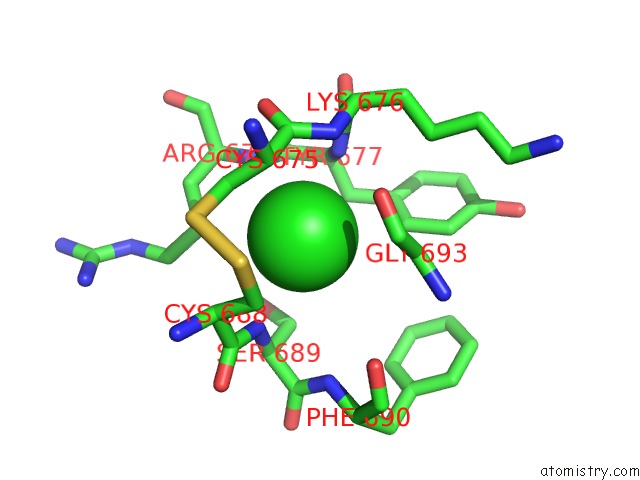
Mono view
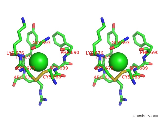
Stereo pair view
Mono view
Stereo pair view
A full contact list of Chlorine with other atoms in the Cl binding
site number 1 of Plexin A2 Extracellular Segment Domains 4-5 (PSI2-IPT2), Resolution 1.36 Angstrom within 5.0Å range:
|
Reference:
Y.Kong,
B.J.Janssen,
T.Malinauskas,
V.R.Vangoor,
C.H.Coles,
R.Kaufmann,
T.Ni,
R.J.Gilbert,
S.Padilla-Parra,
R.J.Pasterkamp,
E.Y.Jones.
Structural Basis For Plexin Activation and Regulation. Neuron V. 91 548 2016.
ISSN: ISSN 1097-4199
PubMed: 27397516
DOI: 10.1016/J.NEURON.2016.06.018
Page generated: Sat Jul 12 04:26:40 2025
ISSN: ISSN 1097-4199
PubMed: 27397516
DOI: 10.1016/J.NEURON.2016.06.018
Last articles
Fe in 2YXOFe in 2YRS
Fe in 2YXC
Fe in 2YNM
Fe in 2YVJ
Fe in 2YP1
Fe in 2YU2
Fe in 2YU1
Fe in 2YQB
Fe in 2YOO