Chlorine »
PDB 5lf2-5ln2 »
5llf »
Chlorine in PDB 5llf: Structure of Polyphosphate Kinase 2 Mutant D117N From Francisella Tularensis with Polyphosphate
Enzymatic activity of Structure of Polyphosphate Kinase 2 Mutant D117N From Francisella Tularensis with Polyphosphate
All present enzymatic activity of Structure of Polyphosphate Kinase 2 Mutant D117N From Francisella Tularensis with Polyphosphate:
2.7.4.1;
2.7.4.1;
Protein crystallography data
The structure of Structure of Polyphosphate Kinase 2 Mutant D117N From Francisella Tularensis with Polyphosphate, PDB code: 5llf
was solved by
P.L.Roach,
A.E.Parnell,
with X-Ray Crystallography technique. A brief refinement statistics is given in the table below:
| Resolution Low / High (Å) | 69.50 / 2.31 |
| Space group | C 2 2 21 |
| Cell size a, b, c (Å), α, β, γ (°) | 73.440, 165.940, 255.300, 90.00, 90.00, 90.00 |
| R / Rfree (%) | 19.8 / 24.7 |
Chlorine Binding Sites:
The binding sites of Chlorine atom in the Structure of Polyphosphate Kinase 2 Mutant D117N From Francisella Tularensis with Polyphosphate
(pdb code 5llf). This binding sites where shown within
5.0 Angstroms radius around Chlorine atom.
In total only one binding site of Chlorine was determined in the Structure of Polyphosphate Kinase 2 Mutant D117N From Francisella Tularensis with Polyphosphate, PDB code: 5llf:
In total only one binding site of Chlorine was determined in the Structure of Polyphosphate Kinase 2 Mutant D117N From Francisella Tularensis with Polyphosphate, PDB code: 5llf:
Chlorine binding site 1 out of 1 in 5llf
Go back to
Chlorine binding site 1 out
of 1 in the Structure of Polyphosphate Kinase 2 Mutant D117N From Francisella Tularensis with Polyphosphate
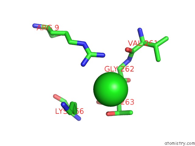
Mono view
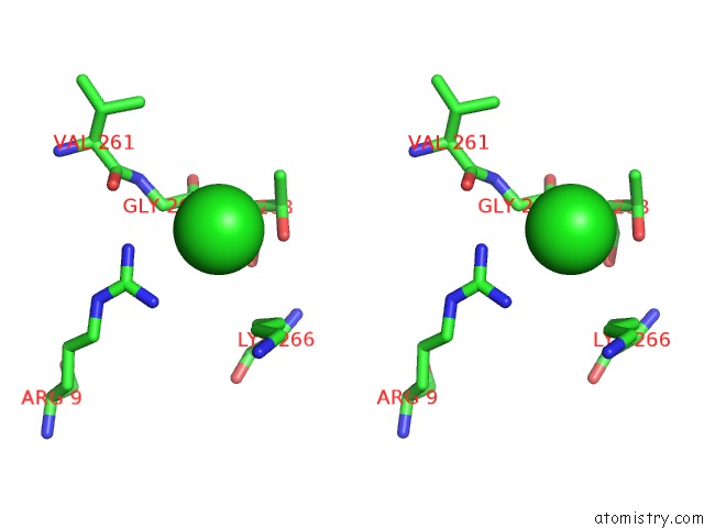
Stereo pair view
Mono view
Stereo pair view
A full contact list of Chlorine with other atoms in the Cl binding
site number 1 of Structure of Polyphosphate Kinase 2 Mutant D117N From Francisella Tularensis with Polyphosphate within 5.0Å range:
|
Reference:
A.E.Parnell,
S.Mordhorst,
F.Kemper,
M.Giurrandino,
J.P.Prince,
N.J.Schwarzer,
A.Hofer,
D.Wohlwend,
H.J.Jessen,
S.Gerhardt,
O.Einsle,
P.C.F.Oyston,
J.N.Andexer,
P.L.Roach.
Substrate Recognition and Mechanism Revealed By Ligand-Bound Polyphosphate Kinase 2 Structures. Proc. Natl. Acad. Sci. V. 115 3350 2018U.S.A..
ISSN: ESSN 1091-6490
PubMed: 29531036
DOI: 10.1073/PNAS.1710741115
Page generated: Fri Jul 26 11:44:17 2024
ISSN: ESSN 1091-6490
PubMed: 29531036
DOI: 10.1073/PNAS.1710741115
Last articles
Zn in 9J0NZn in 9J0O
Zn in 9J0P
Zn in 9FJX
Zn in 9EKB
Zn in 9C0F
Zn in 9CAH
Zn in 9CH0
Zn in 9CH3
Zn in 9CH1