Chlorine »
PDB 5lo9-5lui »
5ltp »
Chlorine in PDB 5ltp: Structure of the Yellow-Green Fluorescent Protein Mneongreen From Branchiostoma Lanceolatum at the Acidic pH 4.5
Protein crystallography data
The structure of Structure of the Yellow-Green Fluorescent Protein Mneongreen From Branchiostoma Lanceolatum at the Acidic pH 4.5, PDB code: 5ltp
was solved by
D.Clavel,
G.Gotthard,
A.Royant,
with X-Ray Crystallography technique. A brief refinement statistics is given in the table below:
| Resolution Low / High (Å) | 46.34 / 1.70 |
| Space group | P 21 21 21 |
| Cell size a, b, c (Å), α, β, γ (°) | 75.870, 127.600, 146.840, 90.00, 90.00, 90.00 |
| R / Rfree (%) | 18.6 / 21.1 |
Chlorine Binding Sites:
The binding sites of Chlorine atom in the Structure of the Yellow-Green Fluorescent Protein Mneongreen From Branchiostoma Lanceolatum at the Acidic pH 4.5
(pdb code 5ltp). This binding sites where shown within
5.0 Angstroms radius around Chlorine atom.
In total 6 binding sites of Chlorine where determined in the Structure of the Yellow-Green Fluorescent Protein Mneongreen From Branchiostoma Lanceolatum at the Acidic pH 4.5, PDB code: 5ltp:
Jump to Chlorine binding site number: 1; 2; 3; 4; 5; 6;
In total 6 binding sites of Chlorine where determined in the Structure of the Yellow-Green Fluorescent Protein Mneongreen From Branchiostoma Lanceolatum at the Acidic pH 4.5, PDB code: 5ltp:
Jump to Chlorine binding site number: 1; 2; 3; 4; 5; 6;
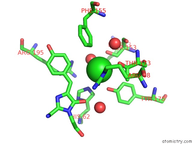
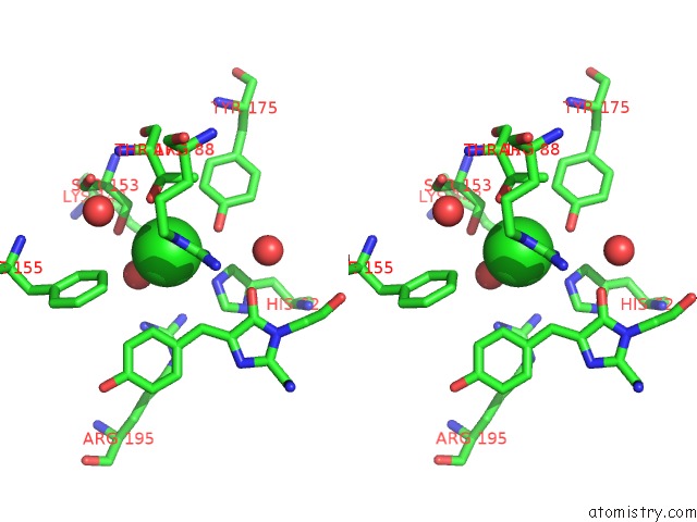
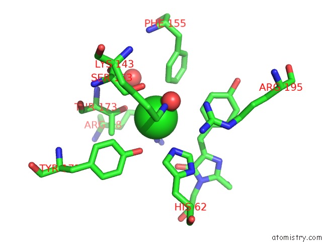
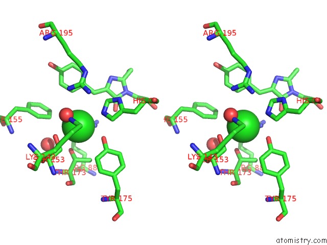
Chlorine binding site 1 out of 6 in 5ltp
Go back to
Chlorine binding site 1 out
of 6 in the Structure of the Yellow-Green Fluorescent Protein Mneongreen From Branchiostoma Lanceolatum at the Acidic pH 4.5
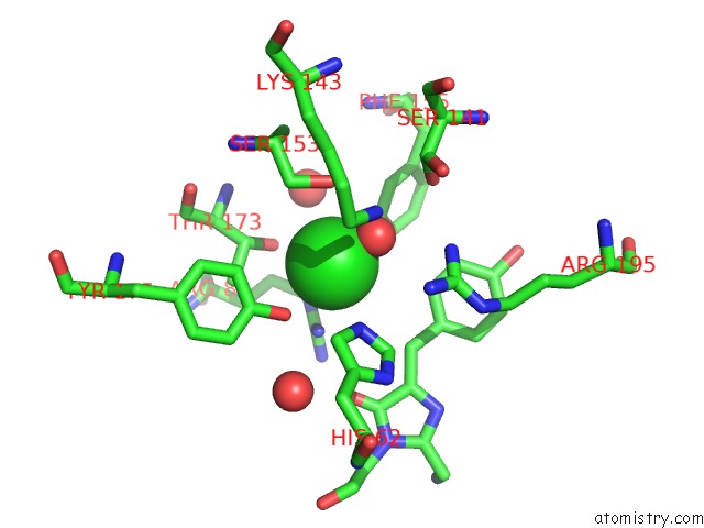
Mono view
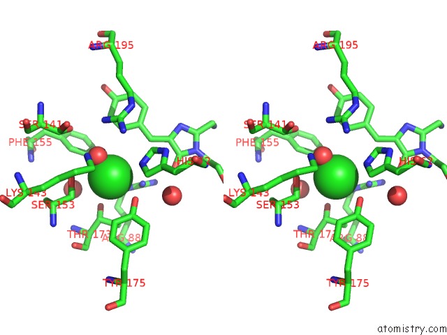
Stereo pair view
Mono view
Stereo pair view
A full contact list of Chlorine with other atoms in the Cl binding
site number 1 of Structure of the Yellow-Green Fluorescent Protein Mneongreen From Branchiostoma Lanceolatum at the Acidic pH 4.5 within 5.0Å range:
|
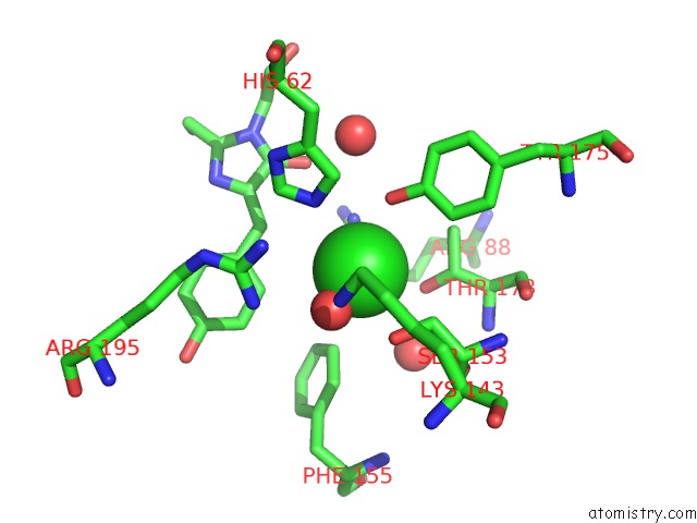
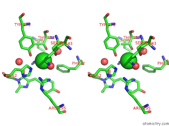
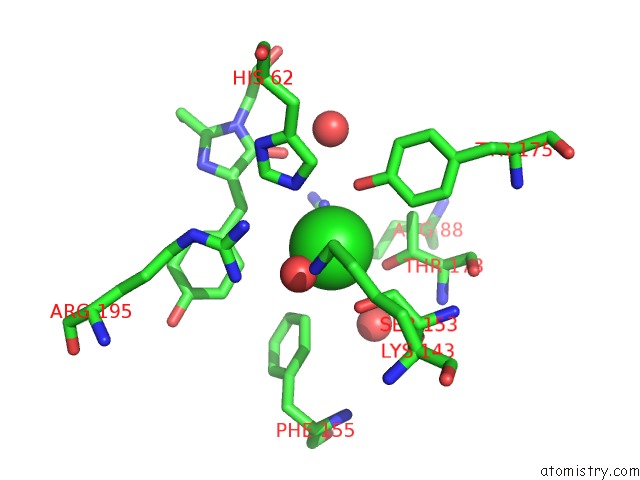
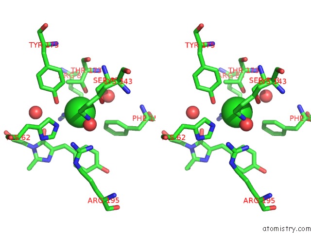
Chlorine binding site 2 out of 6 in 5ltp
Go back to
Chlorine binding site 2 out
of 6 in the Structure of the Yellow-Green Fluorescent Protein Mneongreen From Branchiostoma Lanceolatum at the Acidic pH 4.5
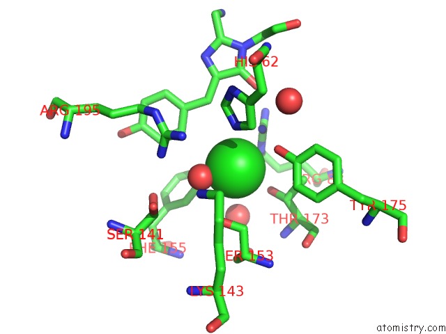
Mono view
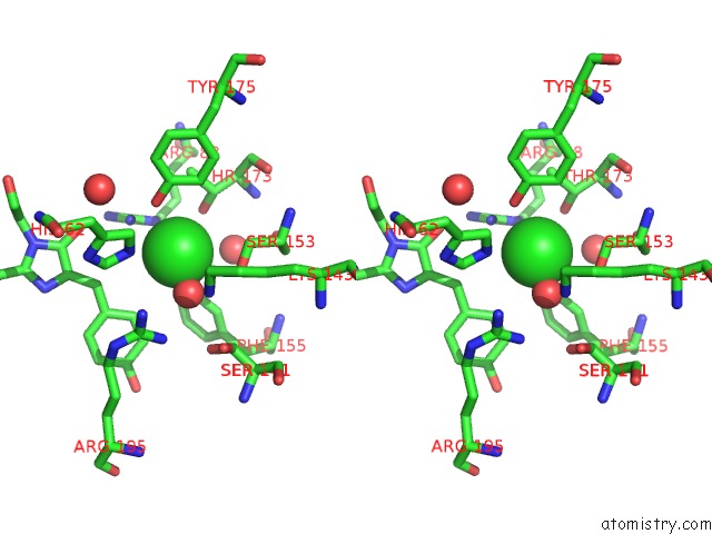
Stereo pair view
Mono view
Stereo pair view
A full contact list of Chlorine with other atoms in the Cl binding
site number 2 of Structure of the Yellow-Green Fluorescent Protein Mneongreen From Branchiostoma Lanceolatum at the Acidic pH 4.5 within 5.0Å range:
|
Chlorine binding site 3 out of 6 in 5ltp
Go back to
Chlorine binding site 3 out
of 6 in the Structure of the Yellow-Green Fluorescent Protein Mneongreen From Branchiostoma Lanceolatum at the Acidic pH 4.5
Mono view
Stereo pair view
Mono view
Stereo pair view
A full contact list of Chlorine with other atoms in the Cl binding
site number 3 of Structure of the Yellow-Green Fluorescent Protein Mneongreen From Branchiostoma Lanceolatum at the Acidic pH 4.5 within 5.0Å range:
|
Chlorine binding site 4 out of 6 in 5ltp
Go back to
Chlorine binding site 4 out
of 6 in the Structure of the Yellow-Green Fluorescent Protein Mneongreen From Branchiostoma Lanceolatum at the Acidic pH 4.5
Mono view
Stereo pair view
Mono view
Stereo pair view
A full contact list of Chlorine with other atoms in the Cl binding
site number 4 of Structure of the Yellow-Green Fluorescent Protein Mneongreen From Branchiostoma Lanceolatum at the Acidic pH 4.5 within 5.0Å range:
|
Chlorine binding site 5 out of 6 in 5ltp
Go back to
Chlorine binding site 5 out
of 6 in the Structure of the Yellow-Green Fluorescent Protein Mneongreen From Branchiostoma Lanceolatum at the Acidic pH 4.5
Mono view
Stereo pair view
Mono view
Stereo pair view
A full contact list of Chlorine with other atoms in the Cl binding
site number 5 of Structure of the Yellow-Green Fluorescent Protein Mneongreen From Branchiostoma Lanceolatum at the Acidic pH 4.5 within 5.0Å range:
|
Chlorine binding site 6 out of 6 in 5ltp
Go back to
Chlorine binding site 6 out
of 6 in the Structure of the Yellow-Green Fluorescent Protein Mneongreen From Branchiostoma Lanceolatum at the Acidic pH 4.5
Mono view
Stereo pair view
Mono view
Stereo pair view
A full contact list of Chlorine with other atoms in the Cl binding
site number 6 of Structure of the Yellow-Green Fluorescent Protein Mneongreen From Branchiostoma Lanceolatum at the Acidic pH 4.5 within 5.0Å range:
|
Reference:
D.Clavel,
G.Gotthard,
D.Von Stetten,
D.De Sanctis,
H.Pasquier,
G.G.Lambert,
N.C.Shaner,
A.Royant.
Structural Analysis of the Bright Monomeric Yellow-Green Fluorescent Protein Mneongreen Obtained By Directed Evolution. Acta Crystallogr D Struct V. 72 1298 2016BIOL.
ISSN: ISSN 2059-7983
PubMed: 27917830
DOI: 10.1107/S2059798316018623
Page generated: Sat Jul 12 05:06:22 2025
ISSN: ISSN 2059-7983
PubMed: 27917830
DOI: 10.1107/S2059798316018623
Last articles
Fe in 2YXOFe in 2YRS
Fe in 2YXC
Fe in 2YNM
Fe in 2YVJ
Fe in 2YP1
Fe in 2YU2
Fe in 2YU1
Fe in 2YQB
Fe in 2YOO