Chlorine »
PDB 5luj-5lzm »
5lwo »
Chlorine in PDB 5lwo: Structure of Spin-Labelled T4 Lysozyme Mutant L115C-R119C-R1 at 100K
Enzymatic activity of Structure of Spin-Labelled T4 Lysozyme Mutant L115C-R119C-R1 at 100K
All present enzymatic activity of Structure of Spin-Labelled T4 Lysozyme Mutant L115C-R119C-R1 at 100K:
3.2.1.17;
3.2.1.17;
Protein crystallography data
The structure of Structure of Spin-Labelled T4 Lysozyme Mutant L115C-R119C-R1 at 100K, PDB code: 5lwo
was solved by
B.Loll,
P.Consentius,
U.Gohlke,
R.Mueller,
M.Kaupp,
U.Heinemann,
M.C.Wahl,
T.Risse,
with X-Ray Crystallography technique. A brief refinement statistics is given in the table below:
| Resolution Low / High (Å) | 18.30 / 1.18 |
| Space group | P 32 2 1 |
| Cell size a, b, c (Å), α, β, γ (°) | 60.170, 60.170, 97.720, 90.00, 90.00, 120.00 |
| R / Rfree (%) | 14.1 / 16.4 |
Other elements in 5lwo:
The structure of Structure of Spin-Labelled T4 Lysozyme Mutant L115C-R119C-R1 at 100K also contains other interesting chemical elements:
| Potassium | (K) | 2 atoms |
Chlorine Binding Sites:
The binding sites of Chlorine atom in the Structure of Spin-Labelled T4 Lysozyme Mutant L115C-R119C-R1 at 100K
(pdb code 5lwo). This binding sites where shown within
5.0 Angstroms radius around Chlorine atom.
In total 4 binding sites of Chlorine where determined in the Structure of Spin-Labelled T4 Lysozyme Mutant L115C-R119C-R1 at 100K, PDB code: 5lwo:
Jump to Chlorine binding site number: 1; 2; 3; 4;
In total 4 binding sites of Chlorine where determined in the Structure of Spin-Labelled T4 Lysozyme Mutant L115C-R119C-R1 at 100K, PDB code: 5lwo:
Jump to Chlorine binding site number: 1; 2; 3; 4;
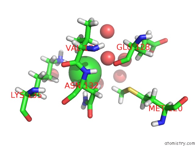
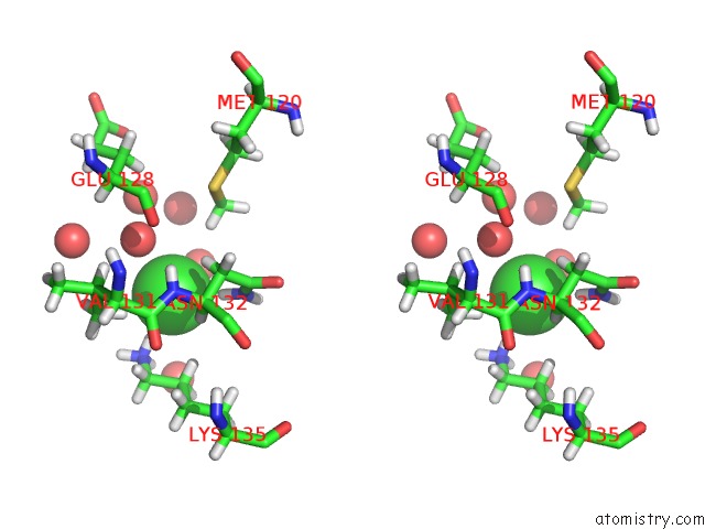
Chlorine binding site 1 out of 4 in 5lwo
Go back to
Chlorine binding site 1 out
of 4 in the Structure of Spin-Labelled T4 Lysozyme Mutant L115C-R119C-R1 at 100K
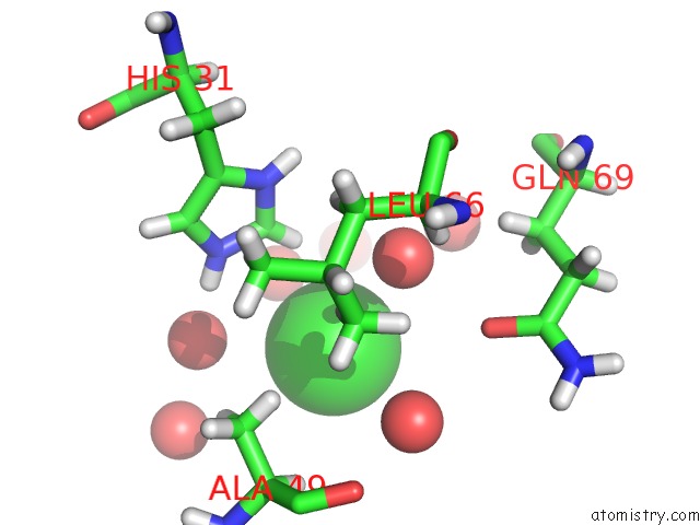
Mono view
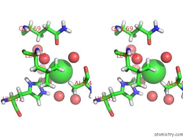
Stereo pair view
Mono view
Stereo pair view
A full contact list of Chlorine with other atoms in the Cl binding
site number 1 of Structure of Spin-Labelled T4 Lysozyme Mutant L115C-R119C-R1 at 100K within 5.0Å range:
|
Chlorine binding site 2 out of 4 in 5lwo
Go back to
Chlorine binding site 2 out
of 4 in the Structure of Spin-Labelled T4 Lysozyme Mutant L115C-R119C-R1 at 100K
Mono view
Stereo pair view
Mono view
Stereo pair view
A full contact list of Chlorine with other atoms in the Cl binding
site number 2 of Structure of Spin-Labelled T4 Lysozyme Mutant L115C-R119C-R1 at 100K within 5.0Å range:
|
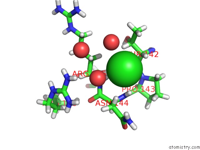
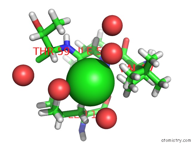
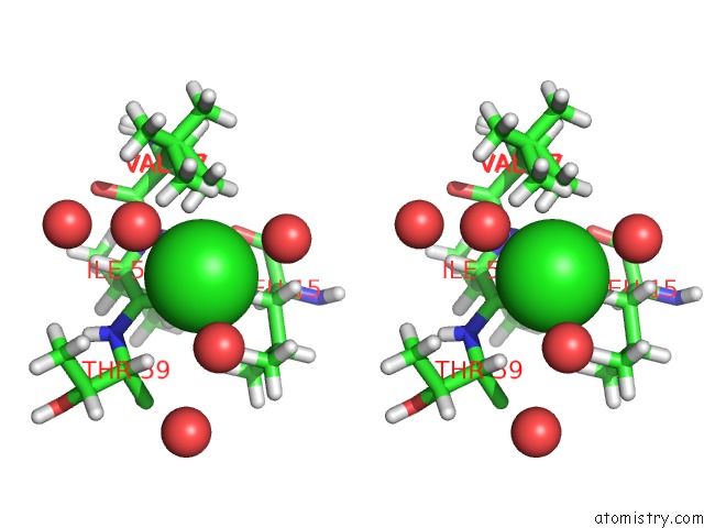
Chlorine binding site 3 out of 4 in 5lwo
Go back to
Chlorine binding site 3 out
of 4 in the Structure of Spin-Labelled T4 Lysozyme Mutant L115C-R119C-R1 at 100K
Mono view
Stereo pair view
Mono view
Stereo pair view
A full contact list of Chlorine with other atoms in the Cl binding
site number 3 of Structure of Spin-Labelled T4 Lysozyme Mutant L115C-R119C-R1 at 100K within 5.0Å range:
|
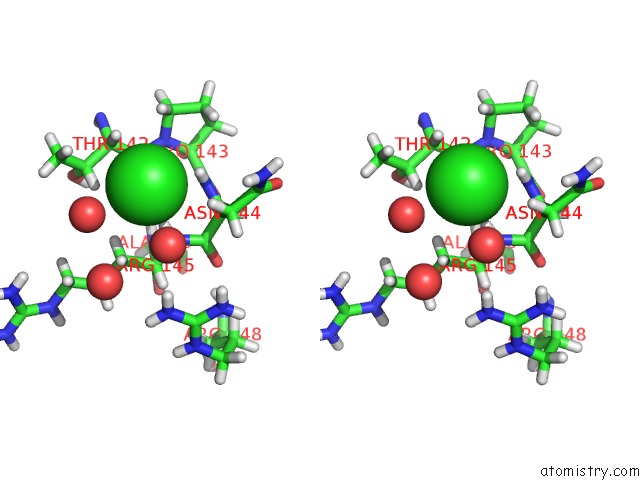
Chlorine binding site 4 out of 4 in 5lwo
Go back to
Chlorine binding site 4 out
of 4 in the Structure of Spin-Labelled T4 Lysozyme Mutant L115C-R119C-R1 at 100K
Mono view
Stereo pair view
Mono view
Stereo pair view
A full contact list of Chlorine with other atoms in the Cl binding
site number 4 of Structure of Spin-Labelled T4 Lysozyme Mutant L115C-R119C-R1 at 100K within 5.0Å range:
|
Reference:
P.Consentius,
B.Loll,
U.Gohlke,
C.Alings,
C.Muller,
R.Muller,
C.Teutloff,
U.Heinemann,
M.Kaupp,
M.C.Wahl,
T.Risse.
Internal Dynamics of the 3-Pyrroline-N-Oxide Ring in Spin-Labeled Proteins. J Phys Chem Lett V. 8 1113 2017.
ISSN: ESSN 1948-7185
PubMed: 28221042
DOI: 10.1021/ACS.JPCLETT.6B02971
Page generated: Sat Jul 12 05:11:25 2025
ISSN: ESSN 1948-7185
PubMed: 28221042
DOI: 10.1021/ACS.JPCLETT.6B02971
Last articles
Fe in 2YXOFe in 2YRS
Fe in 2YXC
Fe in 2YNM
Fe in 2YVJ
Fe in 2YP1
Fe in 2YU2
Fe in 2YU1
Fe in 2YQB
Fe in 2YOO