Chlorine »
PDB 5mtt-5mzc »
5muo »
Chlorine in PDB 5muo: X-Ray Structure of the 2-22' Locally-Closed Mutant of Glic in Complex with Propofol
Protein crystallography data
The structure of X-Ray Structure of the 2-22' Locally-Closed Mutant of Glic in Complex with Propofol, PDB code: 5muo
was solved by
Z.Fourati,
R.R.Ruza,
M.Delarue,
with X-Ray Crystallography technique. A brief refinement statistics is given in the table below:
| Resolution Low / High (Å) | 20.00 / 3.19 |
| Space group | C 1 2 1 |
| Cell size a, b, c (Å), α, β, γ (°) | 180.980, 132.900, 159.920, 90.00, 101.84, 90.00 |
| R / Rfree (%) | 18.9 / 20.4 |
Chlorine Binding Sites:
The binding sites of Chlorine atom in the X-Ray Structure of the 2-22' Locally-Closed Mutant of Glic in Complex with Propofol
(pdb code 5muo). This binding sites where shown within
5.0 Angstroms radius around Chlorine atom.
In total 5 binding sites of Chlorine where determined in the X-Ray Structure of the 2-22' Locally-Closed Mutant of Glic in Complex with Propofol, PDB code: 5muo:
Jump to Chlorine binding site number: 1; 2; 3; 4; 5;
In total 5 binding sites of Chlorine where determined in the X-Ray Structure of the 2-22' Locally-Closed Mutant of Glic in Complex with Propofol, PDB code: 5muo:
Jump to Chlorine binding site number: 1; 2; 3; 4; 5;
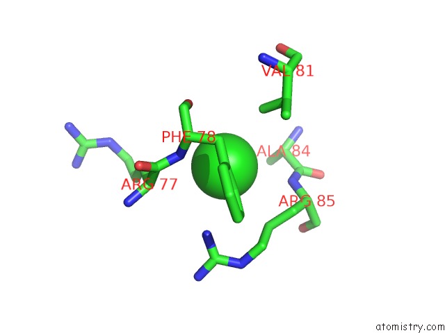
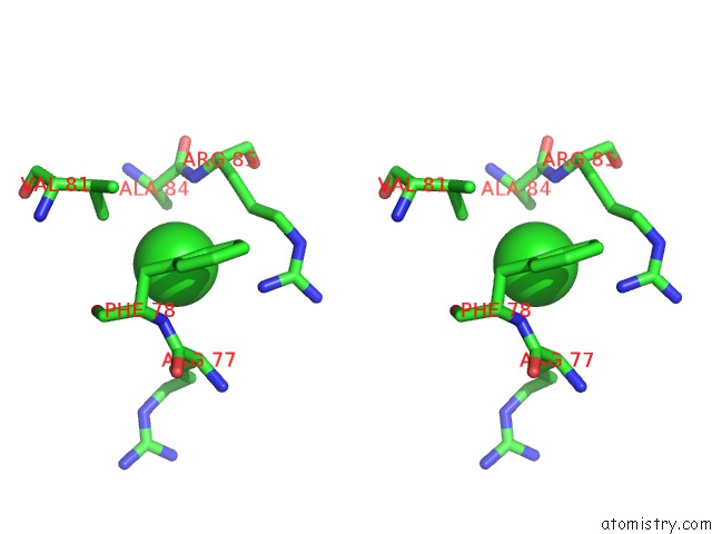
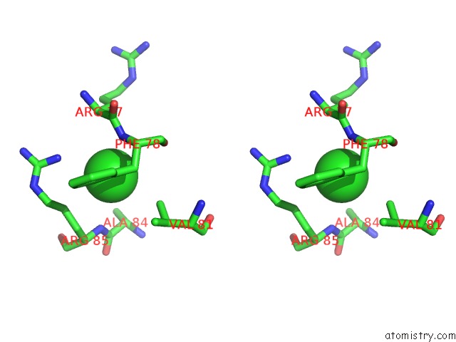
Chlorine binding site 1 out of 5 in 5muo
Go back to
Chlorine binding site 1 out
of 5 in the X-Ray Structure of the 2-22' Locally-Closed Mutant of Glic in Complex with Propofol
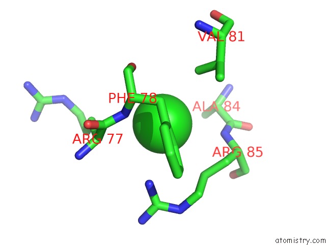
Mono view
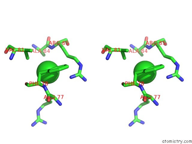
Stereo pair view
Mono view
Stereo pair view
A full contact list of Chlorine with other atoms in the Cl binding
site number 1 of X-Ray Structure of the 2-22' Locally-Closed Mutant of Glic in Complex with Propofol within 5.0Å range:
|
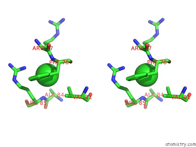
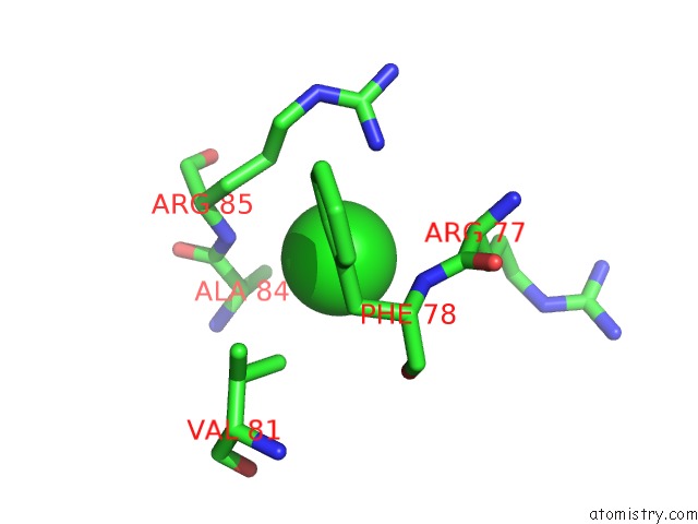
Chlorine binding site 2 out of 5 in 5muo
Go back to
Chlorine binding site 2 out
of 5 in the X-Ray Structure of the 2-22' Locally-Closed Mutant of Glic in Complex with Propofol
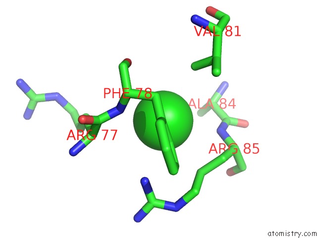
Mono view
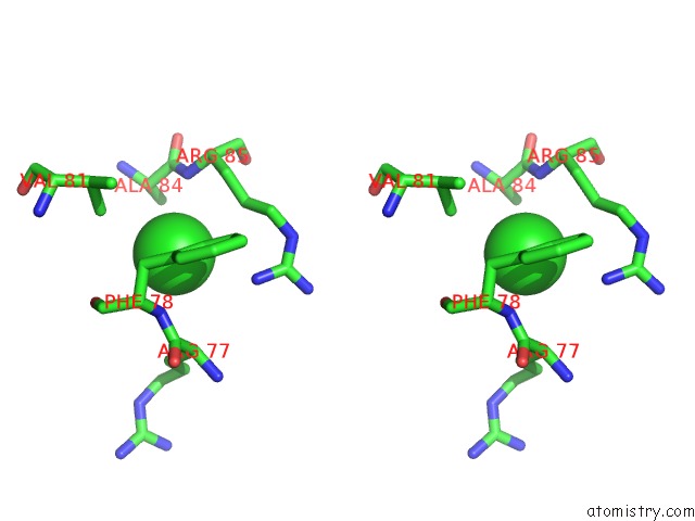
Stereo pair view
Mono view
Stereo pair view
A full contact list of Chlorine with other atoms in the Cl binding
site number 2 of X-Ray Structure of the 2-22' Locally-Closed Mutant of Glic in Complex with Propofol within 5.0Å range:
|
Chlorine binding site 3 out of 5 in 5muo
Go back to
Chlorine binding site 3 out
of 5 in the X-Ray Structure of the 2-22' Locally-Closed Mutant of Glic in Complex with Propofol
Mono view
Stereo pair view
Mono view
Stereo pair view
A full contact list of Chlorine with other atoms in the Cl binding
site number 3 of X-Ray Structure of the 2-22' Locally-Closed Mutant of Glic in Complex with Propofol within 5.0Å range:
|
Chlorine binding site 4 out of 5 in 5muo
Go back to
Chlorine binding site 4 out
of 5 in the X-Ray Structure of the 2-22' Locally-Closed Mutant of Glic in Complex with Propofol

Mono view
Stereo pair view
Mono view
Stereo pair view
A full contact list of Chlorine with other atoms in the Cl binding
site number 4 of X-Ray Structure of the 2-22' Locally-Closed Mutant of Glic in Complex with Propofol within 5.0Å range:
|
Chlorine binding site 5 out of 5 in 5muo
Go back to
Chlorine binding site 5 out
of 5 in the X-Ray Structure of the 2-22' Locally-Closed Mutant of Glic in Complex with Propofol
Mono view
Stereo pair view
Mono view
Stereo pair view
A full contact list of Chlorine with other atoms in the Cl binding
site number 5 of X-Ray Structure of the 2-22' Locally-Closed Mutant of Glic in Complex with Propofol within 5.0Å range:
|
Reference:
Z.Fourati,
R.J.Howard,
S.A.Heusser,
H.Hu,
R.R.Ruza,
L.Sauguet,
E.Lindahl,
M.Delarue.
Structural Basis For A Bimodal Allosteric Mechanism of General Anesthetic Modulation in Pentameric Ligand-Gated Ion Channels. Cell Rep V. 23 993 2018.
ISSN: ESSN 2211-1247
PubMed: 29694907
DOI: 10.1016/J.CELREP.2018.03.108
Page generated: Fri Jul 26 12:56:22 2024
ISSN: ESSN 2211-1247
PubMed: 29694907
DOI: 10.1016/J.CELREP.2018.03.108
Last articles
Zn in 9J0NZn in 9J0O
Zn in 9J0P
Zn in 9FJX
Zn in 9EKB
Zn in 9C0F
Zn in 9CAH
Zn in 9CH0
Zn in 9CH3
Zn in 9CH1