Chlorine »
PDB 5mtt-5mzc »
5mus »
Chlorine in PDB 5mus: Structure of the C-Terminal Domain of A Reptarenavirus L Protein
Protein crystallography data
The structure of Structure of the C-Terminal Domain of A Reptarenavirus L Protein, PDB code: 5mus
was solved by
M.Rosenthal,
N.Gogrefe,
J.Reguera,
D.Vogel,
B.Rauschenberger,
S.Cusack,
S.Gunther,
S.Reindl,
with X-Ray Crystallography technique. A brief refinement statistics is given in the table below:
| Resolution Low / High (Å) | 63.95 / 2.01 |
| Space group | P 21 21 21 |
| Cell size a, b, c (Å), α, β, γ (°) | 76.392, 76.942, 116.920, 90.00, 90.00, 90.00 |
| R / Rfree (%) | 19.8 / 24.2 |
Chlorine Binding Sites:
The binding sites of Chlorine atom in the Structure of the C-Terminal Domain of A Reptarenavirus L Protein
(pdb code 5mus). This binding sites where shown within
5.0 Angstroms radius around Chlorine atom.
In total only one binding site of Chlorine was determined in the Structure of the C-Terminal Domain of A Reptarenavirus L Protein, PDB code: 5mus:
In total only one binding site of Chlorine was determined in the Structure of the C-Terminal Domain of A Reptarenavirus L Protein, PDB code: 5mus:
Chlorine binding site 1 out of 1 in 5mus
Go back to
Chlorine binding site 1 out
of 1 in the Structure of the C-Terminal Domain of A Reptarenavirus L Protein
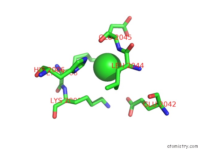
Mono view
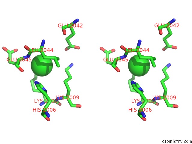
Stereo pair view
Mono view
Stereo pair view
A full contact list of Chlorine with other atoms in the Cl binding
site number 1 of Structure of the C-Terminal Domain of A Reptarenavirus L Protein within 5.0Å range:
|
Reference:
M.Rosenthal,
N.Gogrefe,
D.Vogel,
J.Reguera,
B.Rauschenberger,
S.Cusack,
S.Gunther,
S.Reindl.
Structural Insights Into Reptarenavirus Cap-Snatching Machinery. Plos Pathog. V. 13 06400 2017.
ISSN: ESSN 1553-7374
PubMed: 28505175
DOI: 10.1371/JOURNAL.PPAT.1006400
Page generated: Fri Jul 26 12:56:21 2024
ISSN: ESSN 1553-7374
PubMed: 28505175
DOI: 10.1371/JOURNAL.PPAT.1006400
Last articles
Zn in 9J0NZn in 9J0O
Zn in 9J0P
Zn in 9FJX
Zn in 9EKB
Zn in 9C0F
Zn in 9CAH
Zn in 9CH0
Zn in 9CH3
Zn in 9CH1