Chlorine »
PDB 5mzf-5n6t »
5mzr »
Chlorine in PDB 5mzr: X-Ray Structure of the H235Q Mutant of Glic in Complex with Propofol
Protein crystallography data
The structure of X-Ray Structure of the H235Q Mutant of Glic in Complex with Propofol, PDB code: 5mzr
was solved by
Z.Fourati,
M.Delarue,
with X-Ray Crystallography technique. A brief refinement statistics is given in the table below:
| Resolution Low / High (Å) | 20.00 / 2.65 |
| Space group | C 1 2 1 |
| Cell size a, b, c (Å), α, β, γ (°) | 182.842, 132.989, 161.162, 90.00, 102.83, 90.00 |
| R / Rfree (%) | 19.1 / 21 |
Other elements in 5mzr:
The structure of X-Ray Structure of the H235Q Mutant of Glic in Complex with Propofol also contains other interesting chemical elements:
| Sodium | (Na) | 6 atoms |
Chlorine Binding Sites:
The binding sites of Chlorine atom in the X-Ray Structure of the H235Q Mutant of Glic in Complex with Propofol
(pdb code 5mzr). This binding sites where shown within
5.0 Angstroms radius around Chlorine atom.
In total 7 binding sites of Chlorine where determined in the X-Ray Structure of the H235Q Mutant of Glic in Complex with Propofol, PDB code: 5mzr:
Jump to Chlorine binding site number: 1; 2; 3; 4; 5; 6; 7;
In total 7 binding sites of Chlorine where determined in the X-Ray Structure of the H235Q Mutant of Glic in Complex with Propofol, PDB code: 5mzr:
Jump to Chlorine binding site number: 1; 2; 3; 4; 5; 6; 7;
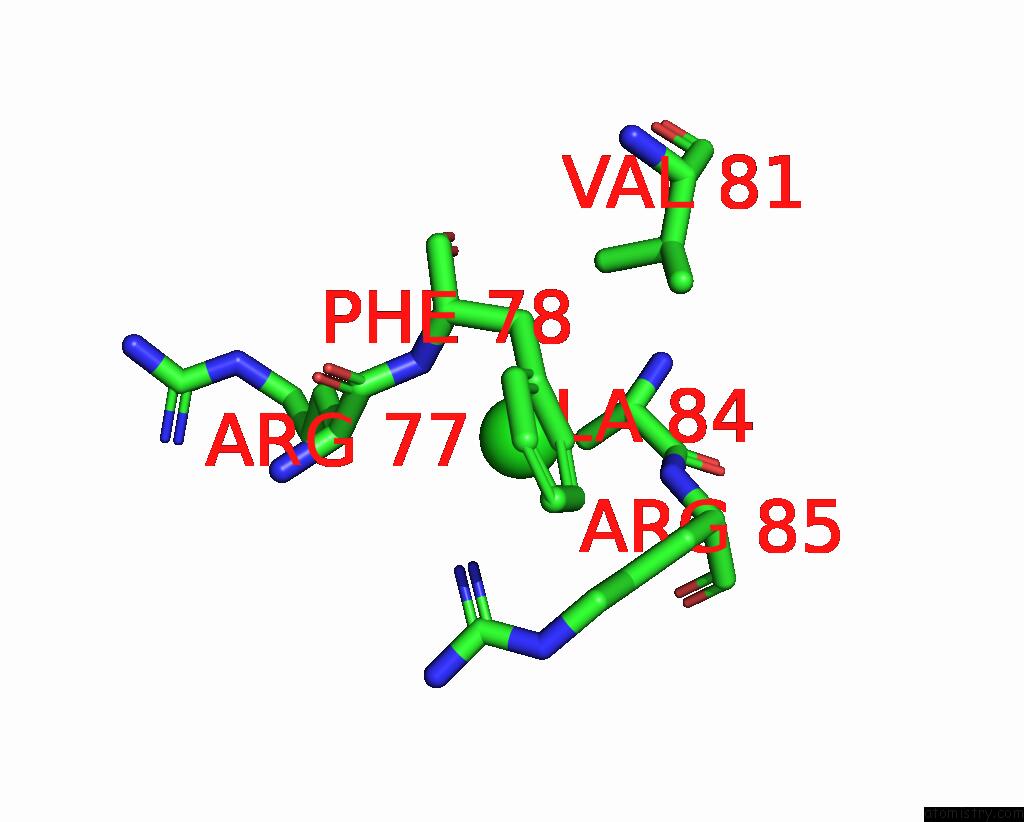
Chlorine binding site 1 out of 7 in 5mzr
Go back to
Chlorine binding site 1 out
of 7 in the X-Ray Structure of the H235Q Mutant of Glic in Complex with Propofol

Mono view

Stereo pair view
Mono view
Stereo pair view
A full contact list of Chlorine with other atoms in the Cl binding
site number 1 of X-Ray Structure of the H235Q Mutant of Glic in Complex with Propofol within 5.0Å range:
|
Chlorine binding site 2 out of 7 in 5mzr
Go back to
Chlorine binding site 2 out
of 7 in the X-Ray Structure of the H235Q Mutant of Glic in Complex with Propofol

Mono view

Stereo pair view
Mono view
Stereo pair view
A full contact list of Chlorine with other atoms in the Cl binding
site number 2 of X-Ray Structure of the H235Q Mutant of Glic in Complex with Propofol within 5.0Å range:
|
Chlorine binding site 3 out of 7 in 5mzr
Go back to
Chlorine binding site 3 out
of 7 in the X-Ray Structure of the H235Q Mutant of Glic in Complex with Propofol

Mono view

Stereo pair view
Mono view
Stereo pair view
A full contact list of Chlorine with other atoms in the Cl binding
site number 3 of X-Ray Structure of the H235Q Mutant of Glic in Complex with Propofol within 5.0Å range:
|
Chlorine binding site 4 out of 7 in 5mzr
Go back to
Chlorine binding site 4 out
of 7 in the X-Ray Structure of the H235Q Mutant of Glic in Complex with Propofol

Mono view

Stereo pair view
Mono view
Stereo pair view
A full contact list of Chlorine with other atoms in the Cl binding
site number 4 of X-Ray Structure of the H235Q Mutant of Glic in Complex with Propofol within 5.0Å range:
|
Chlorine binding site 5 out of 7 in 5mzr
Go back to
Chlorine binding site 5 out
of 7 in the X-Ray Structure of the H235Q Mutant of Glic in Complex with Propofol
Mono view

Stereo pair view
Mono view
Stereo pair view
A full contact list of Chlorine with other atoms in the Cl binding
site number 5 of X-Ray Structure of the H235Q Mutant of Glic in Complex with Propofol within 5.0Å range:
|
Chlorine binding site 6 out of 7 in 5mzr
Go back to
Chlorine binding site 6 out
of 7 in the X-Ray Structure of the H235Q Mutant of Glic in Complex with Propofol

Mono view

Stereo pair view
Mono view
Stereo pair view
A full contact list of Chlorine with other atoms in the Cl binding
site number 6 of X-Ray Structure of the H235Q Mutant of Glic in Complex with Propofol within 5.0Å range:
|
Chlorine binding site 7 out of 7 in 5mzr
Go back to
Chlorine binding site 7 out
of 7 in the X-Ray Structure of the H235Q Mutant of Glic in Complex with Propofol

Mono view

Stereo pair view
Mono view
Stereo pair view
A full contact list of Chlorine with other atoms in the Cl binding
site number 7 of X-Ray Structure of the H235Q Mutant of Glic in Complex with Propofol within 5.0Å range:
|
Reference:
Z.Fourati,
M.Delarue.
X-Ray Structure of the H235Q Mutant of Glic in Complex with Propofol To Be Published.
Page generated: Sat Jul 12 05:46:52 2025
Last articles
F in 4FF6F in 4FIM
F in 4FDO
F in 4FDN
F in 4FC0
F in 4FAT
F in 4F9Y
F in 4FA2
F in 4F9W
F in 4FAD