Chlorine »
PDB 5mzf-5n6t »
5n53 »
Chlorine in PDB 5n53: Crystal Structure of Human 3-Phosphoglycerate Dehydrogenase in Complex with N-(3-Chloro-4-Methoxyphenyl) Acetamide
Enzymatic activity of Crystal Structure of Human 3-Phosphoglycerate Dehydrogenase in Complex with N-(3-Chloro-4-Methoxyphenyl) Acetamide
All present enzymatic activity of Crystal Structure of Human 3-Phosphoglycerate Dehydrogenase in Complex with N-(3-Chloro-4-Methoxyphenyl) Acetamide:
1.1.1.95;
1.1.1.95;
Protein crystallography data
The structure of Crystal Structure of Human 3-Phosphoglycerate Dehydrogenase in Complex with N-(3-Chloro-4-Methoxyphenyl) Acetamide, PDB code: 5n53
was solved by
J.E.Unterlass,
A.Basle,
T.J.Blackburn,
J.Tucker,
C.Cano,
M.E.M.Noble,
N.J.Curtin,
with X-Ray Crystallography technique. A brief refinement statistics is given in the table below:
| Resolution Low / High (Å) | 50.12 / 1.48 |
| Space group | P 1 |
| Cell size a, b, c (Å), α, β, γ (°) | 43.353, 45.323, 55.255, 97.90, 110.06, 106.35 |
| R / Rfree (%) | 18.9 / 24.4 |
Chlorine Binding Sites:
The binding sites of Chlorine atom in the Crystal Structure of Human 3-Phosphoglycerate Dehydrogenase in Complex with N-(3-Chloro-4-Methoxyphenyl) Acetamide
(pdb code 5n53). This binding sites where shown within
5.0 Angstroms radius around Chlorine atom.
In total 2 binding sites of Chlorine where determined in the Crystal Structure of Human 3-Phosphoglycerate Dehydrogenase in Complex with N-(3-Chloro-4-Methoxyphenyl) Acetamide, PDB code: 5n53:
Jump to Chlorine binding site number: 1; 2;
In total 2 binding sites of Chlorine where determined in the Crystal Structure of Human 3-Phosphoglycerate Dehydrogenase in Complex with N-(3-Chloro-4-Methoxyphenyl) Acetamide, PDB code: 5n53:
Jump to Chlorine binding site number: 1; 2;
Chlorine binding site 1 out of 2 in 5n53
Go back to
Chlorine binding site 1 out
of 2 in the Crystal Structure of Human 3-Phosphoglycerate Dehydrogenase in Complex with N-(3-Chloro-4-Methoxyphenyl) Acetamide
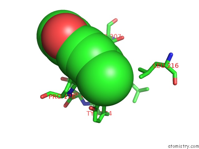
Mono view
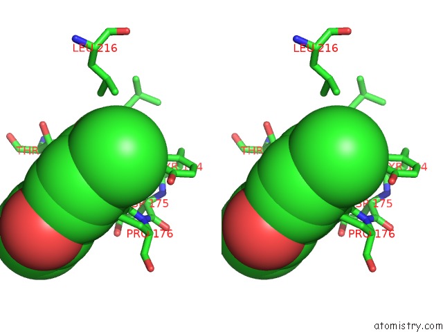
Stereo pair view
Mono view
Stereo pair view
A full contact list of Chlorine with other atoms in the Cl binding
site number 1 of Crystal Structure of Human 3-Phosphoglycerate Dehydrogenase in Complex with N-(3-Chloro-4-Methoxyphenyl) Acetamide within 5.0Å range:
|
Chlorine binding site 2 out of 2 in 5n53
Go back to
Chlorine binding site 2 out
of 2 in the Crystal Structure of Human 3-Phosphoglycerate Dehydrogenase in Complex with N-(3-Chloro-4-Methoxyphenyl) Acetamide
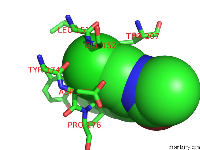
Mono view
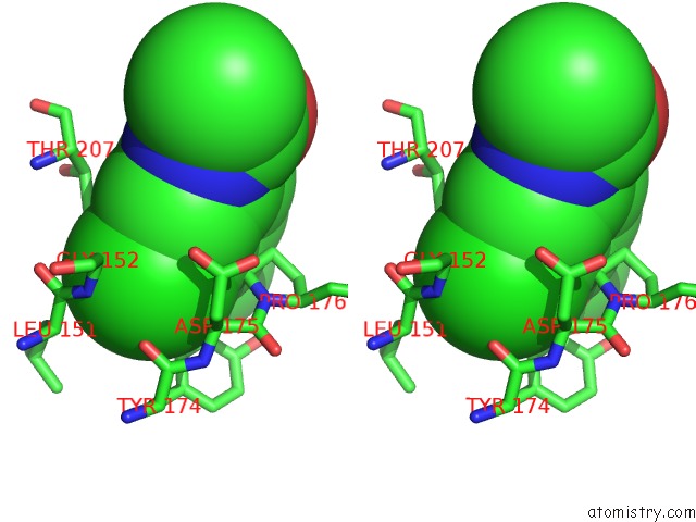
Stereo pair view
Mono view
Stereo pair view
A full contact list of Chlorine with other atoms in the Cl binding
site number 2 of Crystal Structure of Human 3-Phosphoglycerate Dehydrogenase in Complex with N-(3-Chloro-4-Methoxyphenyl) Acetamide within 5.0Å range:
|
Reference:
J.E.Unterlass,
A.Basle,
T.J.Blackburn,
J.Tucker,
C.Cano,
M.E.M.Noble,
N.J.Curtin.
Validating and Enabling Phosphoglycerate Dehydrogenase (Phgdh) As A Target For Fragment-Based Drug Discovery in Phgdh-Amplified Breast Cancer. Oncotarget V. 9 13139 2018.
ISSN: ESSN 1949-2553
PubMed: 29568346
DOI: 10.18632/ONCOTARGET.11487
Page generated: Fri Jul 26 13:12:47 2024
ISSN: ESSN 1949-2553
PubMed: 29568346
DOI: 10.18632/ONCOTARGET.11487
Last articles
Zn in 9J0NZn in 9J0O
Zn in 9J0P
Zn in 9FJX
Zn in 9EKB
Zn in 9C0F
Zn in 9CAH
Zn in 9CH0
Zn in 9CH3
Zn in 9CH1