Chlorine »
PDB 5n75-5ncw »
5na9 »
Chlorine in PDB 5na9: The X-Ray Structure of Bovine Pancreatic Ribonuclease Incubated in the Presence of An Excess of Carboplatin (1:10 Ratio)
Enzymatic activity of The X-Ray Structure of Bovine Pancreatic Ribonuclease Incubated in the Presence of An Excess of Carboplatin (1:10 Ratio)
All present enzymatic activity of The X-Ray Structure of Bovine Pancreatic Ribonuclease Incubated in the Presence of An Excess of Carboplatin (1:10 Ratio):
3.1.27.5;
3.1.27.5;
Protein crystallography data
The structure of The X-Ray Structure of Bovine Pancreatic Ribonuclease Incubated in the Presence of An Excess of Carboplatin (1:10 Ratio), PDB code: 5na9
was solved by
G.Ferraro,
A.Merlino,
with X-Ray Crystallography technique. A brief refinement statistics is given in the table below:
| Resolution Low / High (Å) | 55.57 / 2.07 |
| Space group | P 32 2 1 |
| Cell size a, b, c (Å), α, β, γ (°) | 64.168, 64.168, 64.271, 90.00, 90.00, 120.00 |
| R / Rfree (%) | 17.8 / 24.4 |
Other elements in 5na9:
The structure of The X-Ray Structure of Bovine Pancreatic Ribonuclease Incubated in the Presence of An Excess of Carboplatin (1:10 Ratio) also contains other interesting chemical elements:
| Caesium | (Cs) | 5 atoms |
| Platinum | (Pt) | 2 atoms |
Chlorine Binding Sites:
The binding sites of Chlorine atom in the The X-Ray Structure of Bovine Pancreatic Ribonuclease Incubated in the Presence of An Excess of Carboplatin (1:10 Ratio)
(pdb code 5na9). This binding sites where shown within
5.0 Angstroms radius around Chlorine atom.
In total 9 binding sites of Chlorine where determined in the The X-Ray Structure of Bovine Pancreatic Ribonuclease Incubated in the Presence of An Excess of Carboplatin (1:10 Ratio), PDB code: 5na9:
Jump to Chlorine binding site number: 1; 2; 3; 4; 5; 6; 7; 8; 9;
In total 9 binding sites of Chlorine where determined in the The X-Ray Structure of Bovine Pancreatic Ribonuclease Incubated in the Presence of An Excess of Carboplatin (1:10 Ratio), PDB code: 5na9:
Jump to Chlorine binding site number: 1; 2; 3; 4; 5; 6; 7; 8; 9;
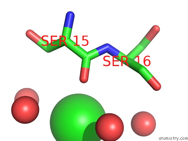
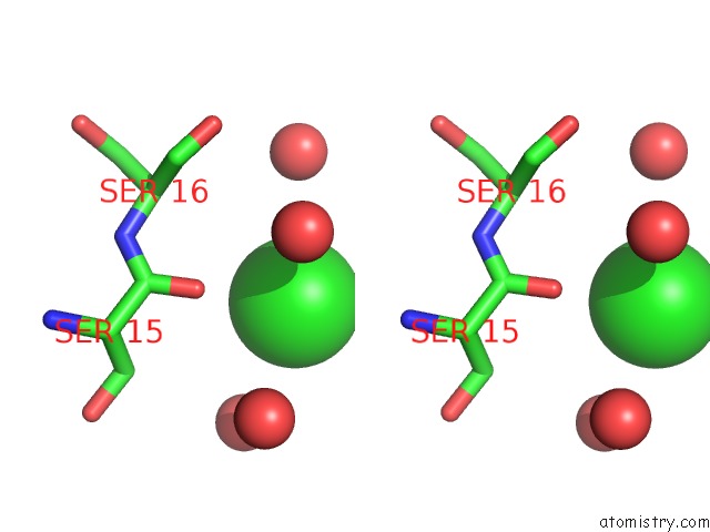
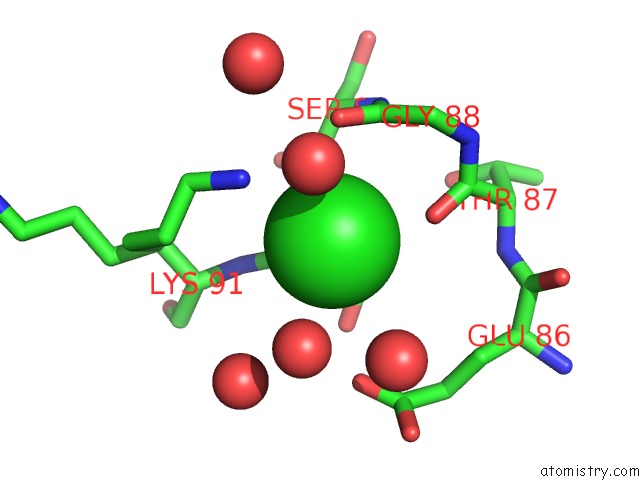
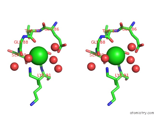
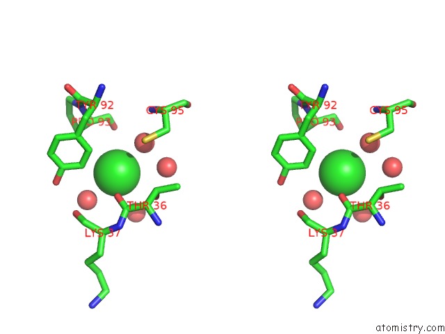
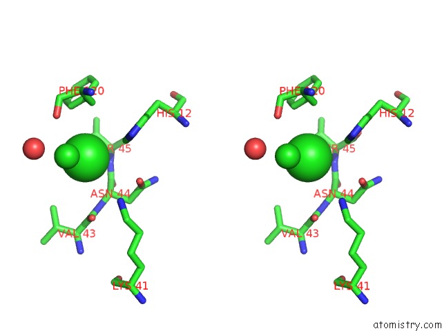
Chlorine binding site 1 out of 9 in 5na9
Go back to
Chlorine binding site 1 out
of 9 in the The X-Ray Structure of Bovine Pancreatic Ribonuclease Incubated in the Presence of An Excess of Carboplatin (1:10 Ratio)
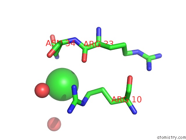
Mono view
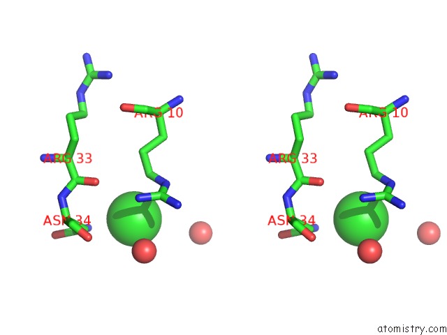
Stereo pair view
Mono view
Stereo pair view
A full contact list of Chlorine with other atoms in the Cl binding
site number 1 of The X-Ray Structure of Bovine Pancreatic Ribonuclease Incubated in the Presence of An Excess of Carboplatin (1:10 Ratio) within 5.0Å range:
|
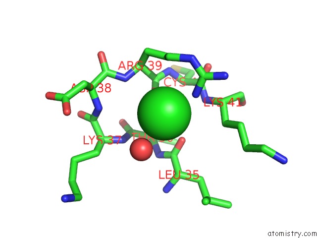
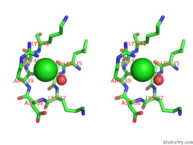
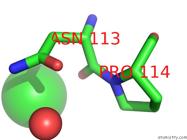
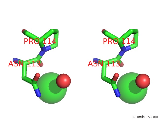
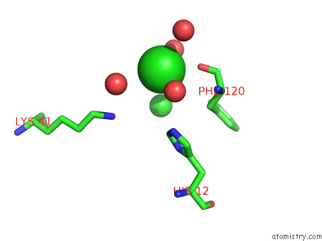
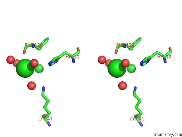
Chlorine binding site 2 out of 9 in 5na9
Go back to
Chlorine binding site 2 out
of 9 in the The X-Ray Structure of Bovine Pancreatic Ribonuclease Incubated in the Presence of An Excess of Carboplatin (1:10 Ratio)
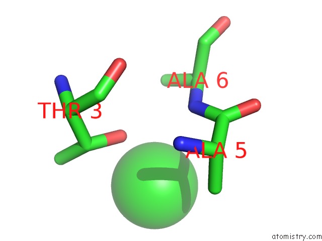
Mono view
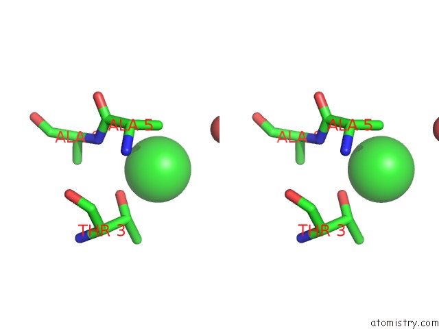
Stereo pair view
Mono view
Stereo pair view
A full contact list of Chlorine with other atoms in the Cl binding
site number 2 of The X-Ray Structure of Bovine Pancreatic Ribonuclease Incubated in the Presence of An Excess of Carboplatin (1:10 Ratio) within 5.0Å range:
|
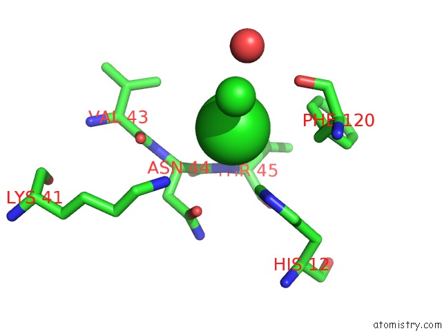
Chlorine binding site 3 out of 9 in 5na9
Go back to
Chlorine binding site 3 out
of 9 in the The X-Ray Structure of Bovine Pancreatic Ribonuclease Incubated in the Presence of An Excess of Carboplatin (1:10 Ratio)
Mono view
Stereo pair view
Mono view
Stereo pair view
A full contact list of Chlorine with other atoms in the Cl binding
site number 3 of The X-Ray Structure of Bovine Pancreatic Ribonuclease Incubated in the Presence of An Excess of Carboplatin (1:10 Ratio) within 5.0Å range:
|
Chlorine binding site 4 out of 9 in 5na9
Go back to
Chlorine binding site 4 out
of 9 in the The X-Ray Structure of Bovine Pancreatic Ribonuclease Incubated in the Presence of An Excess of Carboplatin (1:10 Ratio)
Mono view
Stereo pair view
Mono view
Stereo pair view
A full contact list of Chlorine with other atoms in the Cl binding
site number 4 of The X-Ray Structure of Bovine Pancreatic Ribonuclease Incubated in the Presence of An Excess of Carboplatin (1:10 Ratio) within 5.0Å range:
|
Chlorine binding site 5 out of 9 in 5na9
Go back to
Chlorine binding site 5 out
of 9 in the The X-Ray Structure of Bovine Pancreatic Ribonuclease Incubated in the Presence of An Excess of Carboplatin (1:10 Ratio)
Mono view
Stereo pair view
Mono view
Stereo pair view
A full contact list of Chlorine with other atoms in the Cl binding
site number 5 of The X-Ray Structure of Bovine Pancreatic Ribonuclease Incubated in the Presence of An Excess of Carboplatin (1:10 Ratio) within 5.0Å range:
|
Chlorine binding site 6 out of 9 in 5na9
Go back to
Chlorine binding site 6 out
of 9 in the The X-Ray Structure of Bovine Pancreatic Ribonuclease Incubated in the Presence of An Excess of Carboplatin (1:10 Ratio)
Mono view
Stereo pair view
Mono view
Stereo pair view
A full contact list of Chlorine with other atoms in the Cl binding
site number 6 of The X-Ray Structure of Bovine Pancreatic Ribonuclease Incubated in the Presence of An Excess of Carboplatin (1:10 Ratio) within 5.0Å range:
|
Chlorine binding site 7 out of 9 in 5na9
Go back to
Chlorine binding site 7 out
of 9 in the The X-Ray Structure of Bovine Pancreatic Ribonuclease Incubated in the Presence of An Excess of Carboplatin (1:10 Ratio)

Mono view
Stereo pair view
Mono view
Stereo pair view
A full contact list of Chlorine with other atoms in the Cl binding
site number 7 of The X-Ray Structure of Bovine Pancreatic Ribonuclease Incubated in the Presence of An Excess of Carboplatin (1:10 Ratio) within 5.0Å range:
|
Chlorine binding site 8 out of 9 in 5na9
Go back to
Chlorine binding site 8 out
of 9 in the The X-Ray Structure of Bovine Pancreatic Ribonuclease Incubated in the Presence of An Excess of Carboplatin (1:10 Ratio)
Mono view
Stereo pair view
Mono view
Stereo pair view
A full contact list of Chlorine with other atoms in the Cl binding
site number 8 of The X-Ray Structure of Bovine Pancreatic Ribonuclease Incubated in the Presence of An Excess of Carboplatin (1:10 Ratio) within 5.0Å range:
|
Chlorine binding site 9 out of 9 in 5na9
Go back to
Chlorine binding site 9 out
of 9 in the The X-Ray Structure of Bovine Pancreatic Ribonuclease Incubated in the Presence of An Excess of Carboplatin (1:10 Ratio)
Mono view
Stereo pair view
Mono view
Stereo pair view
A full contact list of Chlorine with other atoms in the Cl binding
site number 9 of The X-Ray Structure of Bovine Pancreatic Ribonuclease Incubated in the Presence of An Excess of Carboplatin (1:10 Ratio) within 5.0Å range:
|
Reference:
D.Picone,
F.Donnarumma,
G.Ferraro,
G.Gotte,
A.Fagagnini,
G.Butera,
M.Donadelli,
A.Merlino.
A Comparison Study on Rnase A Oligomerization Induced By Cisplatin, Carboplatin and Oxaliplatin. J. Inorg. Biochem. V. 173 105 2017.
ISSN: ISSN 1873-3344
PubMed: 28511060
DOI: 10.1016/J.JINORGBIO.2017.05.005
Page generated: Sat Jul 12 05:56:48 2025
ISSN: ISSN 1873-3344
PubMed: 28511060
DOI: 10.1016/J.JINORGBIO.2017.05.005
Last articles
F in 7QN0F in 7QN4
F in 7QN1
F in 7QN2
F in 7QN3
F in 7QMY
F in 7QMX
F in 7QMW
F in 7QMZ
F in 7QMU