Chlorine »
PDB 5nd0-5nkd »
5nf6 »
Chlorine in PDB 5nf6: Structure of GLUK3 Ligand-Binding Domain (S1S2) in Complex with Cip-As at 2.55 A Resolution
Protein crystallography data
The structure of Structure of GLUK3 Ligand-Binding Domain (S1S2) in Complex with Cip-As at 2.55 A Resolution, PDB code: 5nf6
was solved by
K.Frydenvang,
R.Venskutonyte,
T.S.Thorsen,
J.S.Kastrup,
with X-Ray Crystallography technique. A brief refinement statistics is given in the table below:
| Resolution Low / High (Å) | 42.48 / 2.55 |
| Space group | P 2 2 21 |
| Cell size a, b, c (Å), α, β, γ (°) | 130.138, 56.071, 87.516, 90.00, 90.00, 90.00 |
| R / Rfree (%) | 20 / 24.6 |
Other elements in 5nf6:
The structure of Structure of GLUK3 Ligand-Binding Domain (S1S2) in Complex with Cip-As at 2.55 A Resolution also contains other interesting chemical elements:
| Zinc | (Zn) | 6 atoms |
Chlorine Binding Sites:
The binding sites of Chlorine atom in the Structure of GLUK3 Ligand-Binding Domain (S1S2) in Complex with Cip-As at 2.55 A Resolution
(pdb code 5nf6). This binding sites where shown within
5.0 Angstroms radius around Chlorine atom.
In total only one binding site of Chlorine was determined in the Structure of GLUK3 Ligand-Binding Domain (S1S2) in Complex with Cip-As at 2.55 A Resolution, PDB code: 5nf6:
In total only one binding site of Chlorine was determined in the Structure of GLUK3 Ligand-Binding Domain (S1S2) in Complex with Cip-As at 2.55 A Resolution, PDB code: 5nf6:
Chlorine binding site 1 out of 1 in 5nf6
Go back to
Chlorine binding site 1 out
of 1 in the Structure of GLUK3 Ligand-Binding Domain (S1S2) in Complex with Cip-As at 2.55 A Resolution
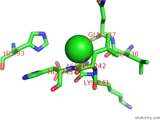
Mono view
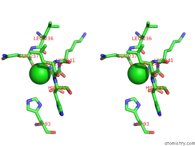
Stereo pair view
Mono view
Stereo pair view
A full contact list of Chlorine with other atoms in the Cl binding
site number 1 of Structure of GLUK3 Ligand-Binding Domain (S1S2) in Complex with Cip-As at 2.55 A Resolution within 5.0Å range:
|
Reference:
S.Mllerud,
A.Pinto,
L.Marconi,
K.Frydenvang,
T.S.Thorsen,
S.Laulumaa,
R.Venskutonyte,
S.Winther,
A.M.C.Moral,
L.Tamborini,
P.Conti,
D.S.Pickering,
J.S.Kastrup.
Structure and Affinity of Two Bicyclic Glutamate Analogues at Ampa and Kainate Receptors. Acs Chem Neurosci V. 8 2056 2017.
ISSN: ESSN 1948-7193
PubMed: 28691798
DOI: 10.1021/ACSCHEMNEURO.7B00201
Page generated: Fri Jul 26 13:31:36 2024
ISSN: ESSN 1948-7193
PubMed: 28691798
DOI: 10.1021/ACSCHEMNEURO.7B00201
Last articles
Zn in 9J0NZn in 9J0O
Zn in 9J0P
Zn in 9FJX
Zn in 9EKB
Zn in 9C0F
Zn in 9CAH
Zn in 9CH0
Zn in 9CH3
Zn in 9CH1