Chlorine »
PDB 5nke-5nry »
5nmt »
Chlorine in PDB 5nmt: Dimer Structure of Sortilin Ectodomain Crystal Form 1, 2.3A
Protein crystallography data
The structure of Dimer Structure of Sortilin Ectodomain Crystal Form 1, 2.3A, PDB code: 5nmt
was solved by
N.O.L.Leloup,
B.J.C.Janssen,
with X-Ray Crystallography technique. A brief refinement statistics is given in the table below:
| Resolution Low / High (Å) | 69.64 / 2.30 |
| Space group | P 21 21 21 |
| Cell size a, b, c (Å), α, β, γ (°) | 97.020, 131.130, 154.630, 90.00, 90.00, 90.00 |
| R / Rfree (%) | 20.5 / 23.4 |
Chlorine Binding Sites:
The binding sites of Chlorine atom in the Dimer Structure of Sortilin Ectodomain Crystal Form 1, 2.3A
(pdb code 5nmt). This binding sites where shown within
5.0 Angstroms radius around Chlorine atom.
In total 2 binding sites of Chlorine where determined in the Dimer Structure of Sortilin Ectodomain Crystal Form 1, 2.3A, PDB code: 5nmt:
Jump to Chlorine binding site number: 1; 2;
In total 2 binding sites of Chlorine where determined in the Dimer Structure of Sortilin Ectodomain Crystal Form 1, 2.3A, PDB code: 5nmt:
Jump to Chlorine binding site number: 1; 2;
Chlorine binding site 1 out of 2 in 5nmt
Go back to
Chlorine binding site 1 out
of 2 in the Dimer Structure of Sortilin Ectodomain Crystal Form 1, 2.3A
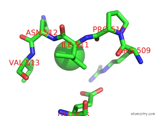
Mono view
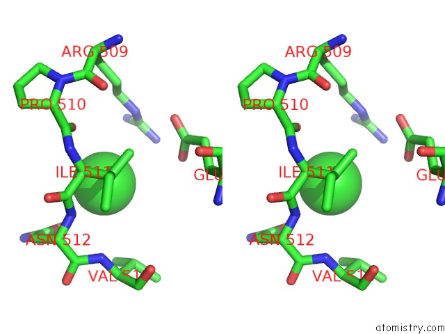
Stereo pair view
Mono view
Stereo pair view
A full contact list of Chlorine with other atoms in the Cl binding
site number 1 of Dimer Structure of Sortilin Ectodomain Crystal Form 1, 2.3A within 5.0Å range:
|
Chlorine binding site 2 out of 2 in 5nmt
Go back to
Chlorine binding site 2 out
of 2 in the Dimer Structure of Sortilin Ectodomain Crystal Form 1, 2.3A
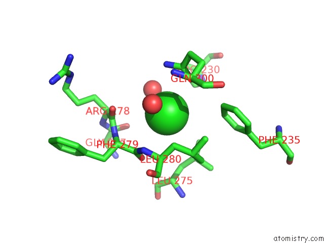
Mono view
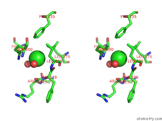
Stereo pair view
Mono view
Stereo pair view
A full contact list of Chlorine with other atoms in the Cl binding
site number 2 of Dimer Structure of Sortilin Ectodomain Crystal Form 1, 2.3A within 5.0Å range:
|
Reference:
N.Leloup,
P.Lossl,
D.H.Meijer,
M.Brennich,
A.J.R.Heck,
D.M.E.Thies-Weesie,
B.J.C.Janssen.
Low pH-Induced Conformational Change and Dimerization of Sortilin Triggers Endocytosed Ligand Release. Nat Commun V. 8 1708 2017.
ISSN: ESSN 2041-1723
PubMed: 29167428
DOI: 10.1038/S41467-017-01485-5
Page generated: Fri Jul 26 13:42:49 2024
ISSN: ESSN 2041-1723
PubMed: 29167428
DOI: 10.1038/S41467-017-01485-5
Last articles
Zn in 9J0NZn in 9J0O
Zn in 9J0P
Zn in 9FJX
Zn in 9EKB
Zn in 9C0F
Zn in 9CAH
Zn in 9CH0
Zn in 9CH3
Zn in 9CH1