Chlorine »
PDB 5nz7-5o5t »
5o5m »
Chlorine in PDB 5o5m: Crystal Structure of the Protein-Kinase A Catalytic Subunit From Criteculus Griseus in Complex with Compounds RKP120 and RKP117
Enzymatic activity of Crystal Structure of the Protein-Kinase A Catalytic Subunit From Criteculus Griseus in Complex with Compounds RKP120 and RKP117
All present enzymatic activity of Crystal Structure of the Protein-Kinase A Catalytic Subunit From Criteculus Griseus in Complex with Compounds RKP120 and RKP117:
2.7.11.11;
2.7.11.11;
Protein crystallography data
The structure of Crystal Structure of the Protein-Kinase A Catalytic Subunit From Criteculus Griseus in Complex with Compounds RKP120 and RKP117, PDB code: 5o5m
was solved by
J.M.Mueller,
A.Heine,
G.Klebe,
with X-Ray Crystallography technique. A brief refinement statistics is given in the table below:
| Resolution Low / High (Å) | 41.92 / 1.58 |
| Space group | P 21 21 21 |
| Cell size a, b, c (Å), α, β, γ (°) | 68.982, 72.633, 76.830, 90.00, 90.00, 90.00 |
| R / Rfree (%) | 14.8 / 18.2 |
Chlorine Binding Sites:
The binding sites of Chlorine atom in the Crystal Structure of the Protein-Kinase A Catalytic Subunit From Criteculus Griseus in Complex with Compounds RKP120 and RKP117
(pdb code 5o5m). This binding sites where shown within
5.0 Angstroms radius around Chlorine atom.
In total only one binding site of Chlorine was determined in the Crystal Structure of the Protein-Kinase A Catalytic Subunit From Criteculus Griseus in Complex with Compounds RKP120 and RKP117, PDB code: 5o5m:
In total only one binding site of Chlorine was determined in the Crystal Structure of the Protein-Kinase A Catalytic Subunit From Criteculus Griseus in Complex with Compounds RKP120 and RKP117, PDB code: 5o5m:
Chlorine binding site 1 out of 1 in 5o5m
Go back to
Chlorine binding site 1 out
of 1 in the Crystal Structure of the Protein-Kinase A Catalytic Subunit From Criteculus Griseus in Complex with Compounds RKP120 and RKP117
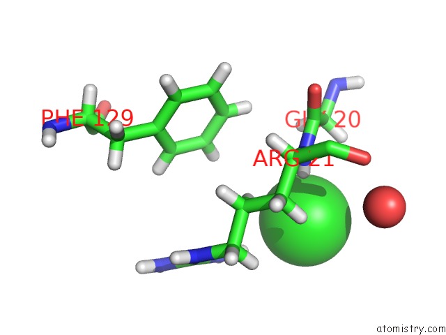
Mono view
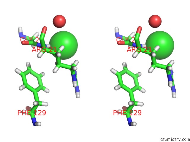
Stereo pair view
Mono view
Stereo pair view
A full contact list of Chlorine with other atoms in the Cl binding
site number 1 of Crystal Structure of the Protein-Kinase A Catalytic Subunit From Criteculus Griseus in Complex with Compounds RKP120 and RKP117 within 5.0Å range:
|
Reference:
J.M.Mueller,
R.Kirschner,
A.Geyer,
G.Klebe.
Conceptional Design of Self-Assembling Bisubstrate-Like Inhibitors of Protein Kinase A Resulting in A Boronic Acid Glutamate Linkage Acs Omega 2019.
ISSN: ESSN 2470-1343
DOI: 10.1021/ACSOMEGA.8B02364
Page generated: Fri Jul 26 14:07:53 2024
ISSN: ESSN 2470-1343
DOI: 10.1021/ACSOMEGA.8B02364
Last articles
Zn in 9J0NZn in 9J0O
Zn in 9J0P
Zn in 9FJX
Zn in 9EKB
Zn in 9C0F
Zn in 9CAH
Zn in 9CH0
Zn in 9CH3
Zn in 9CH1