Chlorine »
PDB 5o5u-5of0 »
5oe2 »
Chlorine in PDB 5oe2: Crystal Structure of the Beta-Lactamase Oxa-245
Enzymatic activity of Crystal Structure of the Beta-Lactamase Oxa-245
All present enzymatic activity of Crystal Structure of the Beta-Lactamase Oxa-245:
3.5.2.6;
3.5.2.6;
Protein crystallography data
The structure of Crystal Structure of the Beta-Lactamase Oxa-245, PDB code: 5oe2
was solved by
B.A.Lund,
H.K.S.Leiros,
with X-Ray Crystallography technique. A brief refinement statistics is given in the table below:
| Resolution Low / High (Å) | 45.26 / 2.20 |
| Space group | P 1 21 1 |
| Cell size a, b, c (Å), α, β, γ (°) | 64.164, 108.720, 83.677, 90.00, 102.39, 90.00 |
| R / Rfree (%) | 19.1 / 22.6 |
Other elements in 5oe2:
The structure of Crystal Structure of the Beta-Lactamase Oxa-245 also contains other interesting chemical elements:
| Sodium | (Na) | 1 atom |
Chlorine Binding Sites:
The binding sites of Chlorine atom in the Crystal Structure of the Beta-Lactamase Oxa-245
(pdb code 5oe2). This binding sites where shown within
5.0 Angstroms radius around Chlorine atom.
In total 2 binding sites of Chlorine where determined in the Crystal Structure of the Beta-Lactamase Oxa-245, PDB code: 5oe2:
Jump to Chlorine binding site number: 1; 2;
In total 2 binding sites of Chlorine where determined in the Crystal Structure of the Beta-Lactamase Oxa-245, PDB code: 5oe2:
Jump to Chlorine binding site number: 1; 2;
Chlorine binding site 1 out of 2 in 5oe2
Go back to
Chlorine binding site 1 out
of 2 in the Crystal Structure of the Beta-Lactamase Oxa-245

Mono view
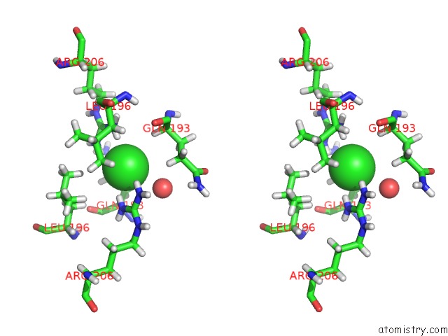
Stereo pair view
Mono view
Stereo pair view
A full contact list of Chlorine with other atoms in the Cl binding
site number 1 of Crystal Structure of the Beta-Lactamase Oxa-245 within 5.0Å range:
|
Chlorine binding site 2 out of 2 in 5oe2
Go back to
Chlorine binding site 2 out
of 2 in the Crystal Structure of the Beta-Lactamase Oxa-245
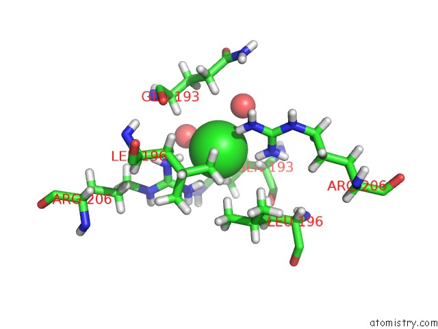
Mono view
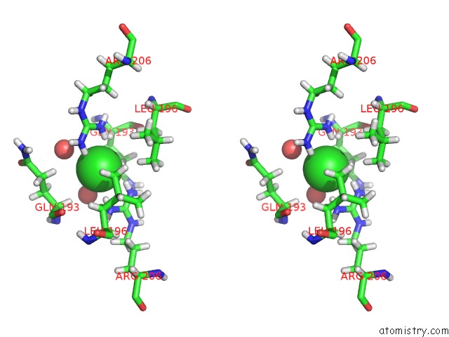
Stereo pair view
Mono view
Stereo pair view
A full contact list of Chlorine with other atoms in the Cl binding
site number 2 of Crystal Structure of the Beta-Lactamase Oxa-245 within 5.0Å range:
|
Reference:
B.A.Lund,
A.M.Thomassen,
T.J.O.Carlsen,
H.K.S.Leiros.
Structure, Activity and Thermostability Investigations of Oxa-163, Oxa-181 and Oxa-245 Using Biochemical Analysis, Crystal Structures and Differential Scanning Calorimetry Analysis. Acta Crystallogr F Struct V. 73 579 2017BIOL Commun.
ISSN: ESSN 2053-230X
PubMed: 28994407
DOI: 10.1107/S2053230X17013838
Page generated: Fri Jul 26 14:26:07 2024
ISSN: ESSN 2053-230X
PubMed: 28994407
DOI: 10.1107/S2053230X17013838
Last articles
Zn in 9J0NZn in 9J0O
Zn in 9J0P
Zn in 9FJX
Zn in 9EKB
Zn in 9C0F
Zn in 9CAH
Zn in 9CH0
Zn in 9CH3
Zn in 9CH1