Chlorine »
PDB 5ofs-5on9 »
5ohq »
Chlorine in PDB 5ohq: Crystal Structure of the KOW6-KOW7 Domain of Human Dsif
Protein crystallography data
The structure of Crystal Structure of the KOW6-KOW7 Domain of Human Dsif, PDB code: 5ohq
was solved by
C.Bernecky,
J.M.Plitzko,
P.Cramer,
with X-Ray Crystallography technique. A brief refinement statistics is given in the table below:
| Resolution Low / High (Å) | 48.37 / 1.10 |
| Space group | C 2 2 21 |
| Cell size a, b, c (Å), α, β, γ (°) | 35.091, 75.657, 96.749, 90.00, 90.00, 90.00 |
| R / Rfree (%) | 12.2 / 14 |
Other elements in 5ohq:
The structure of Crystal Structure of the KOW6-KOW7 Domain of Human Dsif also contains other interesting chemical elements:
| Sodium | (Na) | 1 atom |
Chlorine Binding Sites:
The binding sites of Chlorine atom in the Crystal Structure of the KOW6-KOW7 Domain of Human Dsif
(pdb code 5ohq). This binding sites where shown within
5.0 Angstroms radius around Chlorine atom.
In total only one binding site of Chlorine was determined in the Crystal Structure of the KOW6-KOW7 Domain of Human Dsif, PDB code: 5ohq:
In total only one binding site of Chlorine was determined in the Crystal Structure of the KOW6-KOW7 Domain of Human Dsif, PDB code: 5ohq:
Chlorine binding site 1 out of 1 in 5ohq
Go back to
Chlorine binding site 1 out
of 1 in the Crystal Structure of the KOW6-KOW7 Domain of Human Dsif
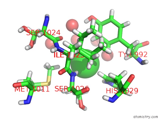
Mono view
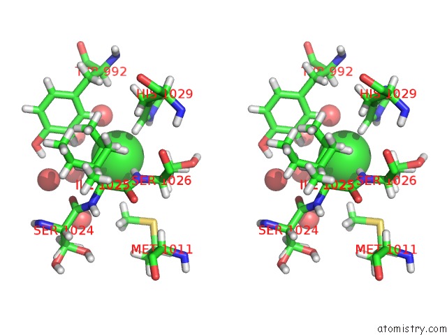
Stereo pair view
Mono view
Stereo pair view
A full contact list of Chlorine with other atoms in the Cl binding
site number 1 of Crystal Structure of the KOW6-KOW7 Domain of Human Dsif within 5.0Å range:
|
Reference:
C.Bernecky,
J.M.Plitzko,
P.Cramer.
Structure of A Transcribing Rna Polymerase II-Dsif Complex Reveals A Multidentate Dna-Rna Clamp. Nat. Struct. Mol. Biol. V. 24 809 2017.
ISSN: ESSN 1545-9985
PubMed: 28892040
DOI: 10.1038/NSMB.3465
Page generated: Fri Jul 26 14:29:42 2024
ISSN: ESSN 1545-9985
PubMed: 28892040
DOI: 10.1038/NSMB.3465
Last articles
Zn in 9J0NZn in 9J0O
Zn in 9J0P
Zn in 9FJX
Zn in 9EKB
Zn in 9C0F
Zn in 9CAH
Zn in 9CH0
Zn in 9CH3
Zn in 9CH1