Chlorine »
PDB 5os5-5oul »
5ou7 »
Chlorine in PDB 5ou7: Crystal Structure of the Glycoprotein VI Loop Truncation Mutant Pavs- Papykn
Protein crystallography data
The structure of Crystal Structure of the Glycoprotein VI Loop Truncation Mutant Pavs- Papykn, PDB code: 5ou7
was solved by
L.J.Feitsma,
E.G.Huizinga,
with X-Ray Crystallography technique. A brief refinement statistics is given in the table below:
| Resolution Low / High (Å) | 113.81 / 1.90 |
| Space group | P 1 21 1 |
| Cell size a, b, c (Å), α, β, γ (°) | 78.650, 44.050, 117.660, 90.00, 104.69, 90.00 |
| R / Rfree (%) | 22.1 / 26 |
Chlorine Binding Sites:
Pages:
>>> Page 1 <<< Page 2, Binding sites: 11 - 14;Binding sites:
The binding sites of Chlorine atom in the Crystal Structure of the Glycoprotein VI Loop Truncation Mutant Pavs- Papykn (pdb code 5ou7). This binding sites where shown within 5.0 Angstroms radius around Chlorine atom.In total 14 binding sites of Chlorine where determined in the Crystal Structure of the Glycoprotein VI Loop Truncation Mutant Pavs- Papykn, PDB code: 5ou7:
Jump to Chlorine binding site number: 1; 2; 3; 4; 5; 6; 7; 8; 9; 10;
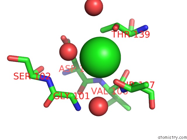
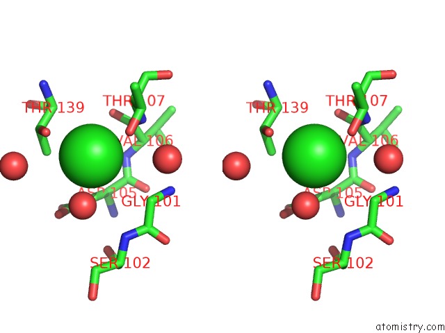
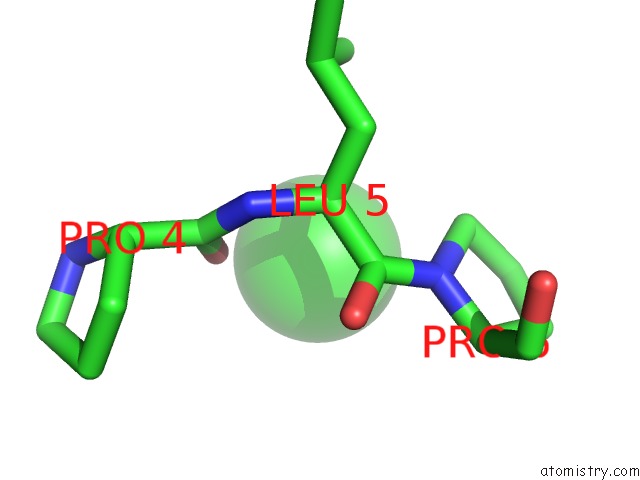
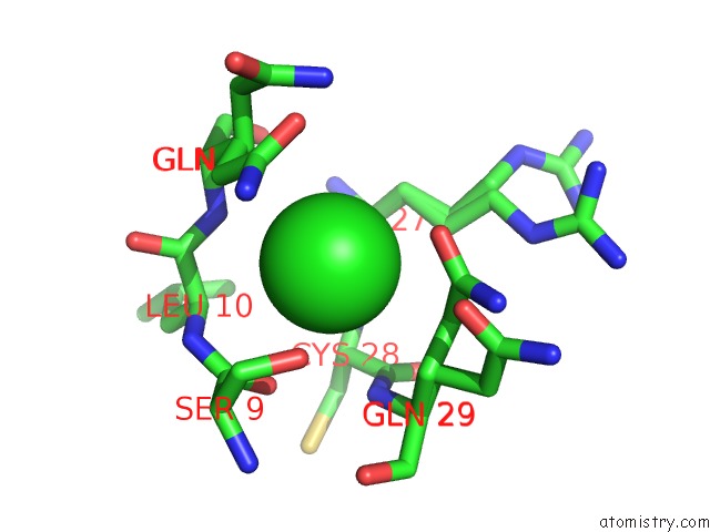
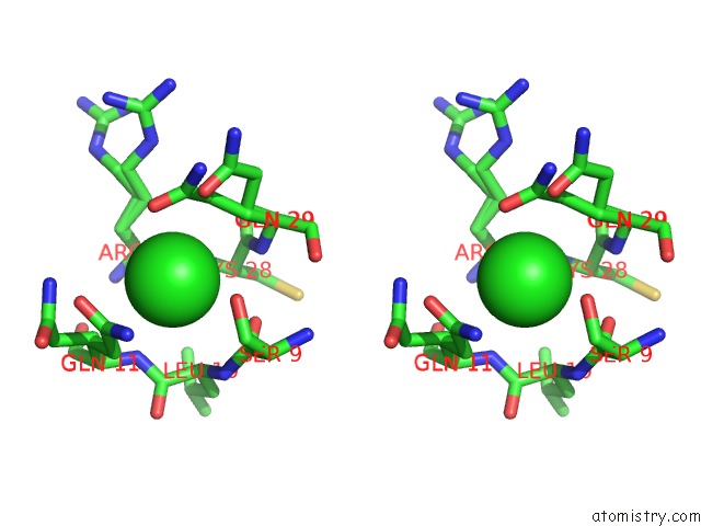
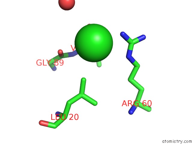
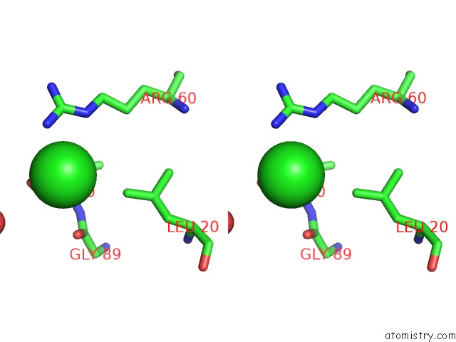
Chlorine binding site 1 out of 14 in 5ou7
Go back to
Chlorine binding site 1 out
of 14 in the Crystal Structure of the Glycoprotein VI Loop Truncation Mutant Pavs- Papykn
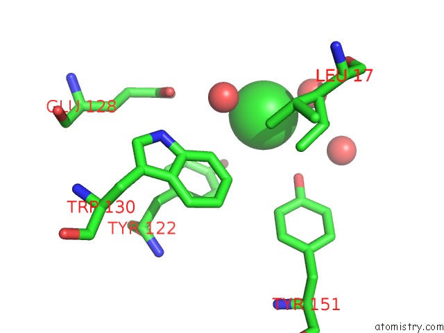
Mono view
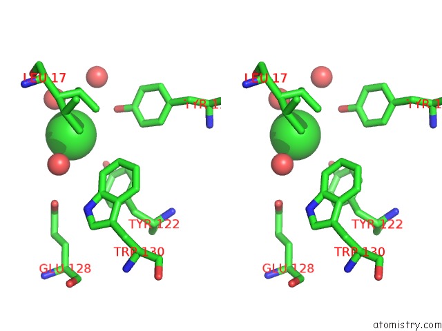
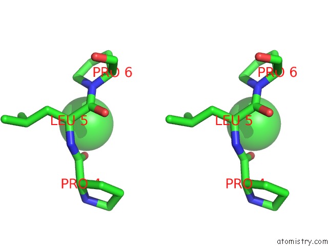
Stereo pair view
Mono view
Stereo pair view
A full contact list of Chlorine with other atoms in the Cl binding
site number 1 of Crystal Structure of the Glycoprotein VI Loop Truncation Mutant Pavs- Papykn within 5.0Å range:
|
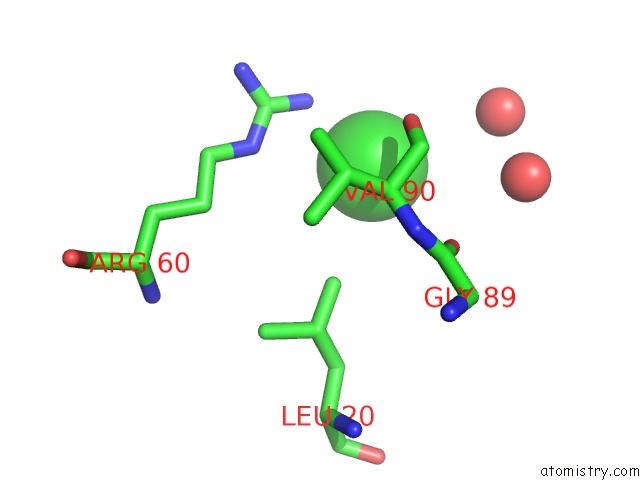
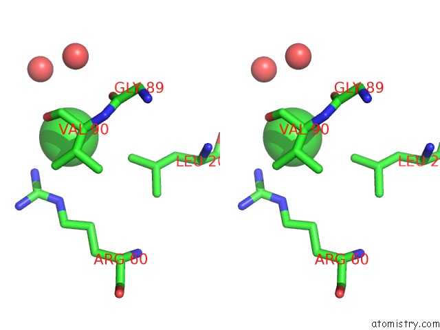
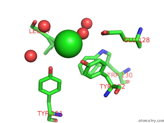
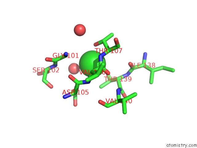
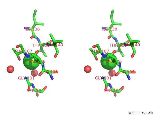
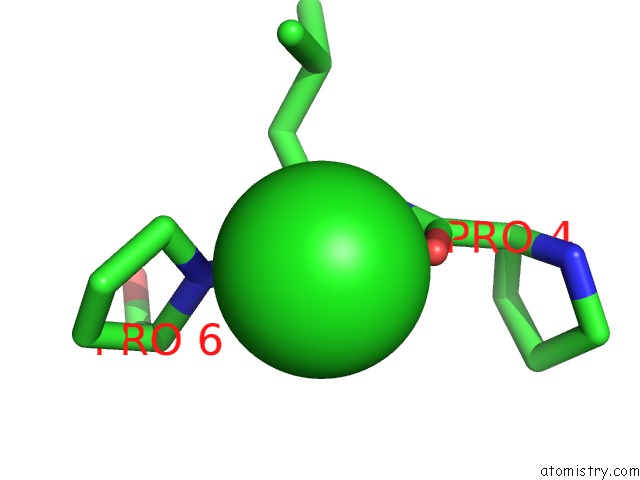
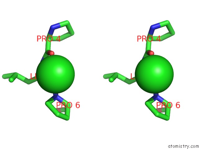
Chlorine binding site 2 out of 14 in 5ou7
Go back to
Chlorine binding site 2 out
of 14 in the Crystal Structure of the Glycoprotein VI Loop Truncation Mutant Pavs- Papykn
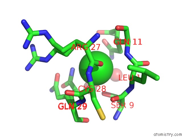
Mono view
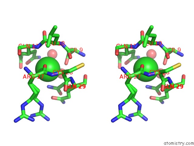
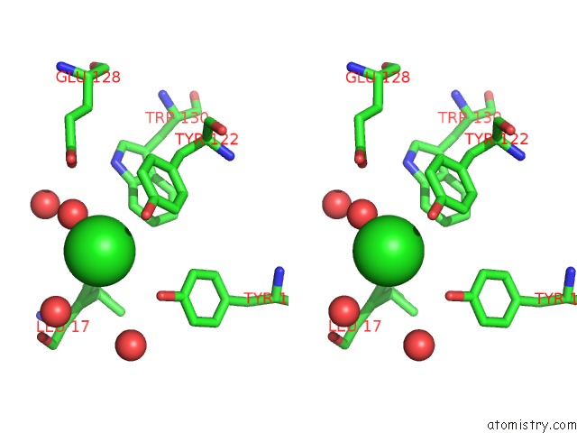
Stereo pair view
Mono view
Stereo pair view
A full contact list of Chlorine with other atoms in the Cl binding
site number 2 of Crystal Structure of the Glycoprotein VI Loop Truncation Mutant Pavs- Papykn within 5.0Å range:
|
Chlorine binding site 3 out of 14 in 5ou7
Go back to
Chlorine binding site 3 out
of 14 in the Crystal Structure of the Glycoprotein VI Loop Truncation Mutant Pavs- Papykn
Mono view
Stereo pair view
Mono view
Stereo pair view
A full contact list of Chlorine with other atoms in the Cl binding
site number 3 of Crystal Structure of the Glycoprotein VI Loop Truncation Mutant Pavs- Papykn within 5.0Å range:
|
Chlorine binding site 4 out of 14 in 5ou7
Go back to
Chlorine binding site 4 out
of 14 in the Crystal Structure of the Glycoprotein VI Loop Truncation Mutant Pavs- Papykn
Mono view
Stereo pair view
Mono view
Stereo pair view
A full contact list of Chlorine with other atoms in the Cl binding
site number 4 of Crystal Structure of the Glycoprotein VI Loop Truncation Mutant Pavs- Papykn within 5.0Å range:
|
Chlorine binding site 5 out of 14 in 5ou7
Go back to
Chlorine binding site 5 out
of 14 in the Crystal Structure of the Glycoprotein VI Loop Truncation Mutant Pavs- Papykn
Mono view
Stereo pair view
Mono view
Stereo pair view
A full contact list of Chlorine with other atoms in the Cl binding
site number 5 of Crystal Structure of the Glycoprotein VI Loop Truncation Mutant Pavs- Papykn within 5.0Å range:
|
Chlorine binding site 6 out of 14 in 5ou7
Go back to
Chlorine binding site 6 out
of 14 in the Crystal Structure of the Glycoprotein VI Loop Truncation Mutant Pavs- Papykn
Mono view
Stereo pair view
Mono view
Stereo pair view
A full contact list of Chlorine with other atoms in the Cl binding
site number 6 of Crystal Structure of the Glycoprotein VI Loop Truncation Mutant Pavs- Papykn within 5.0Å range:
|
Chlorine binding site 7 out of 14 in 5ou7
Go back to
Chlorine binding site 7 out
of 14 in the Crystal Structure of the Glycoprotein VI Loop Truncation Mutant Pavs- Papykn
Mono view
Stereo pair view
Mono view
Stereo pair view
A full contact list of Chlorine with other atoms in the Cl binding
site number 7 of Crystal Structure of the Glycoprotein VI Loop Truncation Mutant Pavs- Papykn within 5.0Å range:
|
Chlorine binding site 8 out of 14 in 5ou7
Go back to
Chlorine binding site 8 out
of 14 in the Crystal Structure of the Glycoprotein VI Loop Truncation Mutant Pavs- Papykn
Mono view
Stereo pair view
Mono view
Stereo pair view
A full contact list of Chlorine with other atoms in the Cl binding
site number 8 of Crystal Structure of the Glycoprotein VI Loop Truncation Mutant Pavs- Papykn within 5.0Å range:
|
Chlorine binding site 9 out of 14 in 5ou7
Go back to
Chlorine binding site 9 out
of 14 in the Crystal Structure of the Glycoprotein VI Loop Truncation Mutant Pavs- Papykn
Mono view
Stereo pair view
Mono view
Stereo pair view
A full contact list of Chlorine with other atoms in the Cl binding
site number 9 of Crystal Structure of the Glycoprotein VI Loop Truncation Mutant Pavs- Papykn within 5.0Å range:
|
Chlorine binding site 10 out of 14 in 5ou7
Go back to
Chlorine binding site 10 out
of 14 in the Crystal Structure of the Glycoprotein VI Loop Truncation Mutant Pavs- Papykn
Mono view
Stereo pair view
Mono view
Stereo pair view
A full contact list of Chlorine with other atoms in the Cl binding
site number 10 of Crystal Structure of the Glycoprotein VI Loop Truncation Mutant Pavs- Papykn within 5.0Å range:
|
Reference:
L.J.Feitsma,
T.H.C.Brondijk,
G.Jarvis,
D.Hagemans,
D.Bihan,
N.Jerah,
M.Versteeg,
R.W.Farndale,
E.G.Huizinga.
Structural Insights Into Collagen-Binding By Platelet Receptor Glycoprotein VI To Be Published.
Page generated: Fri Jul 26 14:52:27 2024
Last articles
Zn in 9JYWZn in 9IR4
Zn in 9IR3
Zn in 9GMX
Zn in 9GMW
Zn in 9JEJ
Zn in 9ERF
Zn in 9ERE
Zn in 9EGV
Zn in 9EGW