Chlorine »
PDB 5qa6-5qca »
5qby »
Chlorine in PDB 5qby: Crystal Structure of Human Cathepsin-S with Bound Ligand
Enzymatic activity of Crystal Structure of Human Cathepsin-S with Bound Ligand
All present enzymatic activity of Crystal Structure of Human Cathepsin-S with Bound Ligand:
3.4.22.27;
3.4.22.27;
Protein crystallography data
The structure of Crystal Structure of Human Cathepsin-S with Bound Ligand, PDB code: 5qby
was solved by
S.D.Bembenek,
M.K.Ameriks,
T.Mirzadegan,
H.Yang,
C.Shao,
S.K.Burley,
with X-Ray Crystallography technique. A brief refinement statistics is given in the table below:
| Resolution Low / High (Å) | 33.87 / 2.25 |
| Space group | C 1 2 1 |
| Cell size a, b, c (Å), α, β, γ (°) | 171.238, 34.376, 103.815, 90.00, 125.10, 90.00 |
| R / Rfree (%) | 17.1 / 20.4 |
Other elements in 5qby:
The structure of Crystal Structure of Human Cathepsin-S with Bound Ligand also contains other interesting chemical elements:
| Fluorine | (F) | 2 atoms |
Chlorine Binding Sites:
The binding sites of Chlorine atom in the Crystal Structure of Human Cathepsin-S with Bound Ligand
(pdb code 5qby). This binding sites where shown within
5.0 Angstroms radius around Chlorine atom.
In total 4 binding sites of Chlorine where determined in the Crystal Structure of Human Cathepsin-S with Bound Ligand, PDB code: 5qby:
Jump to Chlorine binding site number: 1; 2; 3; 4;
In total 4 binding sites of Chlorine where determined in the Crystal Structure of Human Cathepsin-S with Bound Ligand, PDB code: 5qby:
Jump to Chlorine binding site number: 1; 2; 3; 4;
Chlorine binding site 1 out of 4 in 5qby
Go back to
Chlorine binding site 1 out
of 4 in the Crystal Structure of Human Cathepsin-S with Bound Ligand

Mono view
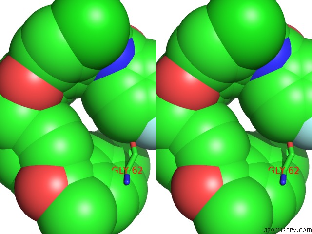
Stereo pair view
Mono view
Stereo pair view
A full contact list of Chlorine with other atoms in the Cl binding
site number 1 of Crystal Structure of Human Cathepsin-S with Bound Ligand within 5.0Å range:
|
Chlorine binding site 2 out of 4 in 5qby
Go back to
Chlorine binding site 2 out
of 4 in the Crystal Structure of Human Cathepsin-S with Bound Ligand
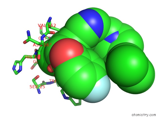
Mono view
Stereo pair view
Mono view
Stereo pair view
A full contact list of Chlorine with other atoms in the Cl binding
site number 2 of Crystal Structure of Human Cathepsin-S with Bound Ligand within 5.0Å range:
|
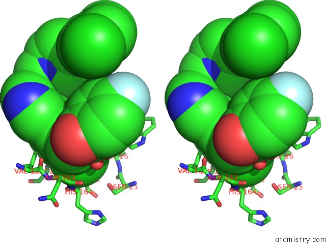
Chlorine binding site 3 out of 4 in 5qby
Go back to
Chlorine binding site 3 out
of 4 in the Crystal Structure of Human Cathepsin-S with Bound Ligand

Mono view

Stereo pair view
Mono view
Stereo pair view
A full contact list of Chlorine with other atoms in the Cl binding
site number 3 of Crystal Structure of Human Cathepsin-S with Bound Ligand within 5.0Å range:
|
Chlorine binding site 4 out of 4 in 5qby
Go back to
Chlorine binding site 4 out
of 4 in the Crystal Structure of Human Cathepsin-S with Bound Ligand
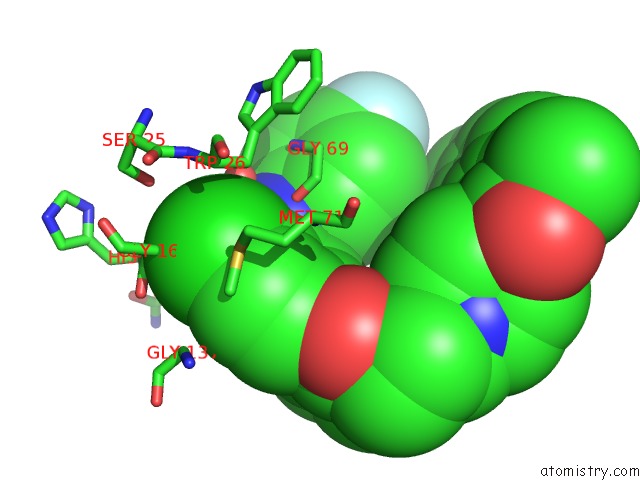
Mono view

Stereo pair view
Mono view
Stereo pair view
A full contact list of Chlorine with other atoms in the Cl binding
site number 4 of Crystal Structure of Human Cathepsin-S with Bound Ligand within 5.0Å range:
|
Reference:
M.K.Ameriks,
S.D.Bembenek,
M.T.Burdett,
I.C.Choong,
J.P.Edwards,
D.Gebauer,
Y.Gu,
L.Karlsson,
H.E.Purkey,
B.L.Staker,
S.Sun,
R.L.Thurmond,
J.Zhu.
Diazinones As P2 Replacements For Pyrazole-Based Cathepsin S Inhibitors Bioorg.Med.Chem.Lett. V. 20 4060 2010.
ISSN: ISSN 0960-894X
PubMed: 20541404
DOI: 10.1016/J.BMCL.2010.05.086
Page generated: Fri Jul 26 15:25:53 2024
ISSN: ISSN 0960-894X
PubMed: 20541404
DOI: 10.1016/J.BMCL.2010.05.086
Last articles
Zn in 9J0NZn in 9J0O
Zn in 9J0P
Zn in 9FJX
Zn in 9EKB
Zn in 9C0F
Zn in 9CAH
Zn in 9CH0
Zn in 9CH3
Zn in 9CH1