Chlorine in PDB 5qch: Crystal Structure of Human Cathepsin-S with Bound Ligand
Enzymatic activity of Crystal Structure of Human Cathepsin-S with Bound Ligand
All present enzymatic activity of Crystal Structure of Human Cathepsin-S with Bound Ligand:
3.4.22.27;
Protein crystallography data
The structure of Crystal Structure of Human Cathepsin-S with Bound Ligand, PDB code: 5qch
was solved by
S.D.Bembenek,
M.K.Ameriks,
T.Mirzadegan,
H.Yang,
C.Shao,
S.K.Burley,
with X-Ray Crystallography technique. A brief refinement statistics is given in the table below:
Resolution Low / High (Å)
|
34.11 /
2.20
|
Space group
|
P 1
|
Cell size a, b, c (Å), α, β, γ (°)
|
54.120,
68.130,
79.020,
88.06,
87.69,
73.08
|
R / Rfree (%)
|
19.9 /
25.1
|
Chlorine Binding Sites:
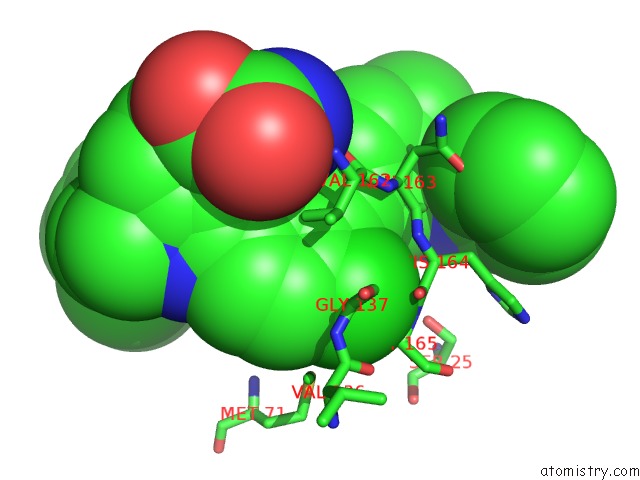
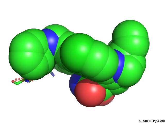
The binding sites of Chlorine atom in the Crystal Structure of Human Cathepsin-S with Bound Ligand
(pdb code 5qch). This binding sites where shown within
5.0 Angstroms radius around Chlorine atom.
In total 8 binding sites of Chlorine where determined in the
Crystal Structure of Human Cathepsin-S with Bound Ligand, PDB code: 5qch:
Jump to Chlorine binding site number:
1;
2;
3;
4;
5;
6;
7;
8;
Chlorine binding site 1 out
of 8 in 5qch
Go back to
Chlorine Binding Sites List in 5qch
Chlorine binding site 1 out
of 8 in the Crystal Structure of Human Cathepsin-S with Bound Ligand
 Mono view
 Stereo pair view
|
A full contact list of Chlorine with other atoms in the Cl binding
site number 1 of Crystal Structure of Human Cathepsin-S with Bound Ligand within 5.0Å range:
probe
|
atom
|
residue
|
distance (Å)
|
B
|
Occ
|
A:Cl802
b:22.3
occ:1.00
|
CL3
|
A:BJS802
|
0.0
|
22.3
|
1.0
|
C74
|
A:BJS802
|
1.7
|
21.3
|
1.0
|
C73
|
A:BJS802
|
2.7
|
19.0
|
1.0
|
C75
|
A:BJS802
|
2.7
|
19.0
|
1.0
|
NH2
|
A:ARG141
|
3.0
|
34.8
|
1.0
|
C85
|
A:BJS802
|
3.1
|
17.4
|
1.0
|
O
|
A:HOH1004
|
3.4
|
33.0
|
1.0
|
C89
|
A:BJS802
|
3.7
|
16.7
|
1.0
|
N87
|
A:BJS802
|
3.9
|
15.4
|
1.0
|
C72
|
A:BJS802
|
4.0
|
19.5
|
1.0
|
C76
|
A:BJS802
|
4.0
|
18.8
|
1.0
|
O
|
A:HOH1069
|
4.0
|
17.7
|
1.0
|
CZ
|
A:ARG141
|
4.2
|
49.4
|
1.0
|
C70
|
A:BJS802
|
4.5
|
18.8
|
1.0
|
NE
|
A:ARG141
|
4.8
|
39.7
|
1.0
|
|
Chlorine binding site 2 out
of 8 in 5qch
Go back to
Chlorine Binding Sites List in 5qch
Chlorine binding site 2 out
of 8 in the Crystal Structure of Human Cathepsin-S with Bound Ligand
 Mono view
 Stereo pair view
|
A full contact list of Chlorine with other atoms in the Cl binding
site number 2 of Crystal Structure of Human Cathepsin-S with Bound Ligand within 5.0Å range:
probe
|
atom
|
residue
|
distance (Å)
|
B
|
Occ
|
A:Cl802
b:23.1
occ:1.00
|
CL4
|
A:BJS802
|
0.0
|
23.1
|
1.0
|
C55
|
A:BJS802
|
1.7
|
22.5
|
1.0
|
C56
|
A:BJS802
|
2.7
|
22.7
|
1.0
|
C54
|
A:BJS802
|
2.7
|
19.2
|
1.0
|
C66
|
A:BJS802
|
3.1
|
18.6
|
1.0
|
N
|
A:GLY165
|
3.4
|
11.0
|
1.0
|
CA
|
A:GLY165
|
3.5
|
10.1
|
1.0
|
C
|
A:HIS164
|
3.6
|
14.8
|
1.0
|
C68
|
A:BJS802
|
3.9
|
18.8
|
1.0
|
O
|
A:HIS164
|
4.0
|
13.0
|
1.0
|
C57
|
A:BJS802
|
4.0
|
22.4
|
1.0
|
C53
|
A:BJS802
|
4.0
|
21.1
|
1.0
|
CA
|
A:GLY137
|
4.0
|
9.2
|
1.0
|
SD
|
A:MET71
|
4.1
|
16.4
|
1.0
|
N
|
A:HIS164
|
4.2
|
9.7
|
1.0
|
CE
|
A:MET71
|
4.2
|
12.8
|
1.0
|
CA
|
A:HIS164
|
4.3
|
9.2
|
1.0
|
C51
|
A:BJS802
|
4.5
|
20.9
|
1.0
|
O
|
A:HOH1068
|
4.8
|
31.0
|
1.0
|
O
|
A:HOH1093
|
4.8
|
24.9
|
1.0
|
O
|
A:GLY69
|
4.8
|
16.6
|
1.0
|
N
|
A:GLY137
|
4.8
|
10.0
|
1.0
|
C
|
A:ASN163
|
4.9
|
13.8
|
1.0
|
CG2
|
A:VAL162
|
4.9
|
13.8
|
1.0
|
O
|
A:VAL136
|
4.9
|
14.2
|
1.0
|
OG
|
A:SER25
|
4.9
|
21.5
|
1.0
|
|
Chlorine binding site 3 out
of 8 in 5qch
Go back to
Chlorine Binding Sites List in 5qch
Chlorine binding site 3 out
of 8 in the Crystal Structure of Human Cathepsin-S with Bound Ligand
 Mono view
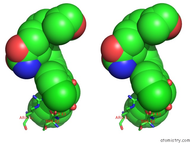
 Stereo pair view
|
A full contact list of Chlorine with other atoms in the Cl binding
site number 3 of Crystal Structure of Human Cathepsin-S with Bound Ligand within 5.0Å range:
probe
|
atom
|
residue
|
distance (Å)
|
B
|
Occ
|
B:Cl803
b:21.1
occ:1.00
|
CL3
|
B:BJS803
|
0.0
|
21.1
|
1.0
|
C74
|
B:BJS803
|
1.7
|
19.1
|
1.0
|
C73
|
B:BJS803
|
2.7
|
15.6
|
1.0
|
C75
|
B:BJS803
|
2.7
|
17.1
|
1.0
|
NH2
|
B:ARG141
|
3.0
|
30.9
|
1.0
|
C85
|
B:BJS803
|
3.1
|
11.9
|
1.0
|
C89
|
B:BJS803
|
3.7
|
8.9
|
1.0
|
O
|
B:HOH1120
|
3.7
|
23.1
|
1.0
|
O
|
D:THR42
|
3.8
|
16.6
|
1.0
|
O
|
D:HOH1153
|
3.9
|
15.1
|
1.0
|
N87
|
B:BJS803
|
3.9
|
9.1
|
1.0
|
C72
|
B:BJS803
|
4.0
|
14.4
|
1.0
|
C76
|
B:BJS803
|
4.0
|
16.8
|
1.0
|
CA
|
D:GLY43
|
4.0
|
14.3
|
1.0
|
CZ
|
B:ARG141
|
4.2
|
41.8
|
1.0
|
C70
|
B:BJS803
|
4.5
|
15.7
|
1.0
|
C
|
D:THR42
|
4.6
|
15.8
|
1.0
|
N
|
D:GLY43
|
4.7
|
13.9
|
1.0
|
C
|
D:GLY43
|
4.8
|
18.2
|
1.0
|
NE
|
B:ARG141
|
4.9
|
31.0
|
1.0
|
O
|
D:GLY43
|
4.9
|
17.9
|
1.0
|
|
Chlorine binding site 4 out
of 8 in 5qch
Go back to
Chlorine Binding Sites List in 5qch
Chlorine binding site 4 out
of 8 in the Crystal Structure of Human Cathepsin-S with Bound Ligand
 Mono view
 Stereo pair view
|
A full contact list of Chlorine with other atoms in the Cl binding
site number 4 of Crystal Structure of Human Cathepsin-S with Bound Ligand within 5.0Å range:
probe
|
atom
|
residue
|
distance (Å)
|
B
|
Occ
|
B:Cl803
b:21.7
occ:1.00
|
CL4
|
B:BJS803
|
0.0
|
21.7
|
1.0
|
C55
|
B:BJS803
|
1.7
|
19.8
|
1.0
|
C56
|
B:BJS803
|
2.7
|
17.7
|
1.0
|
C54
|
B:BJS803
|
2.7
|
16.8
|
1.0
|
C66
|
B:BJS803
|
3.1
|
17.2
|
1.0
|
N
|
B:GLY165
|
3.4
|
12.4
|
1.0
|
CA
|
B:GLY165
|
3.5
|
11.7
|
1.0
|
C
|
B:HIS164
|
3.7
|
15.3
|
1.0
|
C68
|
B:BJS803
|
3.8
|
17.0
|
1.0
|
C53
|
B:BJS803
|
4.0
|
15.9
|
1.0
|
C57
|
B:BJS803
|
4.0
|
16.9
|
1.0
|
O
|
B:HIS164
|
4.0
|
14.4
|
1.0
|
CA
|
B:GLY137
|
4.1
|
11.6
|
1.0
|
SD
|
B:MET71
|
4.1
|
16.3
|
1.0
|
N
|
B:HIS164
|
4.2
|
11.1
|
1.0
|
CE
|
B:MET71
|
4.2
|
12.6
|
1.0
|
CA
|
B:HIS164
|
4.3
|
10.2
|
1.0
|
C51
|
B:BJS803
|
4.5
|
15.0
|
1.0
|
O
|
B:HOH1058
|
4.5
|
15.4
|
1.0
|
O
|
B:GLY69
|
4.8
|
18.0
|
1.0
|
C
|
B:ASN163
|
4.8
|
15.1
|
1.0
|
N
|
B:GLY137
|
4.9
|
11.6
|
1.0
|
CG2
|
B:VAL162
|
4.9
|
13.3
|
1.0
|
OG
|
B:SER25
|
4.9
|
11.7
|
1.0
|
O
|
B:VAL136
|
4.9
|
13.8
|
1.0
|
|
Chlorine binding site 5 out
of 8 in 5qch
Go back to
Chlorine Binding Sites List in 5qch
Chlorine binding site 5 out
of 8 in the Crystal Structure of Human Cathepsin-S with Bound Ligand
 Mono view
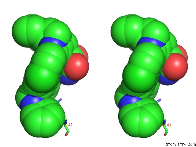
 Stereo pair view
|
A full contact list of Chlorine with other atoms in the Cl binding
site number 5 of Crystal Structure of Human Cathepsin-S with Bound Ligand within 5.0Å range:
probe
|
atom
|
residue
|
distance (Å)
|
B
|
Occ
|
C:Cl802
b:23.6
occ:1.00
|
CL3
|
C:BJS802
|
0.0
|
23.6
|
1.0
|
C74
|
C:BJS802
|
1.7
|
20.0
|
1.0
|
C73
|
C:BJS802
|
2.7
|
16.9
|
1.0
|
C75
|
C:BJS802
|
2.7
|
15.3
|
1.0
|
NH2
|
C:ARG141
|
3.0
|
34.1
|
1.0
|
C85
|
C:BJS802
|
3.0
|
12.1
|
1.0
|
C89
|
C:BJS802
|
3.7
|
13.4
|
1.0
|
N87
|
C:BJS802
|
3.9
|
11.4
|
1.0
|
C72
|
C:BJS802
|
4.0
|
15.6
|
1.0
|
C76
|
C:BJS802
|
4.0
|
13.2
|
1.0
|
O
|
C:HOH1076
|
4.0
|
45.0
|
1.0
|
CZ
|
C:ARG141
|
4.2
|
46.5
|
1.0
|
C70
|
C:BJS802
|
4.5
|
15.7
|
1.0
|
O
|
C:HOH1158
|
4.8
|
38.1
|
1.0
|
NE
|
C:ARG141
|
4.9
|
39.1
|
1.0
|
|
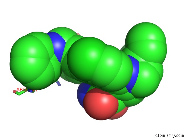
Chlorine binding site 6 out
of 8 in 5qch
Go back to
Chlorine Binding Sites List in 5qch
Chlorine binding site 6 out
of 8 in the Crystal Structure of Human Cathepsin-S with Bound Ligand
 Mono view
 Stereo pair view
|
A full contact list of Chlorine with other atoms in the Cl binding
site number 6 of Crystal Structure of Human Cathepsin-S with Bound Ligand within 5.0Å range:
probe
|
atom
|
residue
|
distance (Å)
|
B
|
Occ
|
C:Cl802
b:20.8
occ:1.00
|
CL4
|
C:BJS802
|
0.0
|
20.8
|
1.0
|
C55
|
C:BJS802
|
1.7
|
21.3
|
1.0
|
C56
|
C:BJS802
|
2.7
|
20.2
|
1.0
|
C54
|
C:BJS802
|
2.7
|
18.9
|
1.0
|
C66
|
C:BJS802
|
3.1
|
17.6
|
1.0
|
N
|
C:GLY165
|
3.3
|
13.8
|
1.0
|
CA
|
C:GLY165
|
3.5
|
12.8
|
1.0
|
C
|
C:HIS164
|
3.6
|
16.0
|
1.0
|
C68
|
C:BJS802
|
3.9
|
17.1
|
1.0
|
O
|
C:HIS164
|
4.0
|
15.0
|
1.0
|
C57
|
C:BJS802
|
4.0
|
18.4
|
1.0
|
C53
|
C:BJS802
|
4.0
|
19.8
|
1.0
|
CA
|
C:GLY137
|
4.1
|
14.3
|
1.0
|
SD
|
C:MET71
|
4.1
|
21.1
|
1.0
|
N
|
C:HIS164
|
4.2
|
11.6
|
1.0
|
CE
|
C:MET71
|
4.3
|
16.7
|
1.0
|
CA
|
C:HIS164
|
4.3
|
10.0
|
1.0
|
O
|
C:HOH1043
|
4.4
|
12.4
|
1.0
|
C51
|
C:BJS802
|
4.5
|
19.2
|
1.0
|
O
|
C:GLY69
|
4.8
|
17.7
|
1.0
|
C
|
C:ASN163
|
4.8
|
15.4
|
1.0
|
N
|
C:GLY137
|
4.9
|
13.7
|
1.0
|
CG2
|
C:VAL162
|
4.9
|
11.6
|
1.0
|
O
|
C:VAL136
|
4.9
|
16.2
|
1.0
|
O
|
C:HOH992
|
4.9
|
18.8
|
1.0
|
OG
|
C:SER25
|
4.9
|
21.2
|
1.0
|
|
Chlorine binding site 7 out
of 8 in 5qch
Go back to
Chlorine Binding Sites List in 5qch
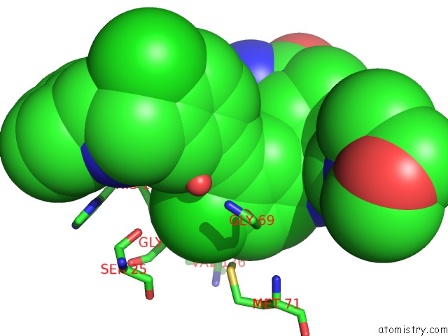
Chlorine binding site 7 out
of 8 in the Crystal Structure of Human Cathepsin-S with Bound Ligand
 Mono view
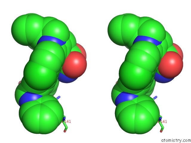
 Stereo pair view
|
A full contact list of Chlorine with other atoms in the Cl binding
site number 7 of Crystal Structure of Human Cathepsin-S with Bound Ligand within 5.0Å range:
probe
|
atom
|
residue
|
distance (Å)
|
B
|
Occ
|
D:Cl803
b:21.9
occ:1.00
|
CL3
|
D:BJS803
|
0.0
|
21.9
|
1.0
|
C74
|
D:BJS803
|
1.8
|
19.9
|
1.0
|
C73
|
D:BJS803
|
2.7
|
17.4
|
1.0
|
C75
|
D:BJS803
|
2.7
|
16.3
|
1.0
|
NH2
|
D:ARG141
|
3.0
|
33.2
|
1.0
|
C85
|
D:BJS803
|
3.1
|
14.8
|
1.0
|
C89
|
D:BJS803
|
3.7
|
15.3
|
1.0
|
O
|
D:HOH1120
|
3.7
|
26.4
|
1.0
|
N87
|
D:BJS803
|
3.9
|
13.4
|
1.0
|
C72
|
D:BJS803
|
4.0
|
14.5
|
1.0
|
C76
|
D:BJS803
|
4.0
|
14.5
|
1.0
|
CZ
|
D:ARG141
|
4.2
|
40.6
|
1.0
|
C70
|
D:BJS803
|
4.5
|
16.2
|
1.0
|
O
|
D:HOH1139
|
4.6
|
51.6
|
1.0
|
NE
|
D:ARG141
|
4.9
|
29.3
|
1.0
|
O
|
D:HOH1121
|
4.9
|
32.8
|
1.0
|
|
Chlorine binding site 8 out
of 8 in 5qch
Go back to
Chlorine Binding Sites List in 5qch
Chlorine binding site 8 out
of 8 in the Crystal Structure of Human Cathepsin-S with Bound Ligand
 Mono view
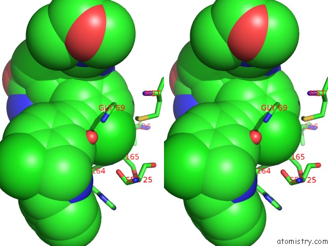
 Stereo pair view
|
A full contact list of Chlorine with other atoms in the Cl binding
site number 8 of Crystal Structure of Human Cathepsin-S with Bound Ligand within 5.0Å range:
probe
|
atom
|
residue
|
distance (Å)
|
B
|
Occ
|
D:Cl803
b:20.3
occ:1.00
|
CL4
|
D:BJS803
|
0.0
|
20.3
|
1.0
|
C55
|
D:BJS803
|
1.8
|
19.8
|
1.0
|
C56
|
D:BJS803
|
2.7
|
18.0
|
1.0
|
C54
|
D:BJS803
|
2.8
|
16.8
|
1.0
|
C66
|
D:BJS803
|
3.1
|
16.9
|
1.0
|
N
|
D:GLY165
|
3.3
|
12.4
|
1.0
|
CA
|
D:GLY165
|
3.5
|
12.0
|
1.0
|
C
|
D:HIS164
|
3.6
|
13.9
|
1.0
|
C68
|
D:BJS803
|
3.9
|
17.0
|
1.0
|
O
|
D:HIS164
|
4.0
|
12.2
|
1.0
|
C57
|
D:BJS803
|
4.0
|
16.9
|
1.0
|
C53
|
D:BJS803
|
4.0
|
15.5
|
1.0
|
CA
|
D:GLY137
|
4.1
|
13.6
|
1.0
|
SD
|
D:MET71
|
4.1
|
17.3
|
1.0
|
N
|
D:HIS164
|
4.2
|
8.9
|
1.0
|
CE
|
D:MET71
|
4.3
|
13.0
|
1.0
|
CA
|
D:HIS164
|
4.3
|
7.7
|
1.0
|
O
|
D:HOH1082
|
4.5
|
21.1
|
1.0
|
C51
|
D:BJS803
|
4.6
|
16.0
|
1.0
|
N
|
D:GLY137
|
4.8
|
13.5
|
1.0
|
C
|
D:ASN163
|
4.9
|
13.4
|
1.0
|
O
|
D:GLY69
|
4.9
|
15.6
|
1.0
|
O
|
D:VAL136
|
4.9
|
15.8
|
1.0
|
OG
|
D:SER25
|
4.9
|
20.0
|
1.0
|
CG2
|
D:VAL162
|
4.9
|
14.4
|
1.0
|
C
|
D:GLY165
|
5.0
|
16.1
|
1.0
|
|
Reference:
S.D.Bembenek,
M.K.Ameriks,
T.Mirzadegan,
H.Yang,
C.Shao,
S.K.Burley.
Crystal Structure of Human Cathepsin-S with Bound Ligand To Be Published.
Page generated: Fri Jul 26 15:27:40 2024
|