Chlorine »
PDB 5syw-5t6k »
5t5x »
Chlorine in PDB 5t5x: High Resolution Structure of Mouse Cryptochrome 1
Protein crystallography data
The structure of High Resolution Structure of Mouse Cryptochrome 1, PDB code: 5t5x
was solved by
A.K.Michael,
S.Tripathi,
C.L.Partch,
with X-Ray Crystallography technique. A brief refinement statistics is given in the table below:
| Resolution Low / High (Å) | 48.43 / 1.84 |
| Space group | P 1 |
| Cell size a, b, c (Å), α, β, γ (°) | 43.630, 51.035, 54.175, 71.62, 84.12, 88.89 |
| R / Rfree (%) | 16.8 / 23.2 |
Chlorine Binding Sites:
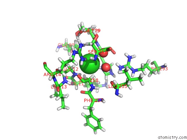
The binding sites of Chlorine atom in the High Resolution Structure of Mouse Cryptochrome 1
(pdb code 5t5x). This binding sites where shown within
5.0 Angstroms radius around Chlorine atom.
In total only one binding site of Chlorine was determined in the High Resolution Structure of Mouse Cryptochrome 1, PDB code: 5t5x:
In total only one binding site of Chlorine was determined in the High Resolution Structure of Mouse Cryptochrome 1, PDB code: 5t5x:
Chlorine binding site 1 out of 1 in 5t5x
Go back to
Chlorine binding site 1 out
of 1 in the High Resolution Structure of Mouse Cryptochrome 1
Mono view

Stereo pair view
Mono view
Stereo pair view
A full contact list of Chlorine with other atoms in the Cl binding
site number 1 of High Resolution Structure of Mouse Cryptochrome 1 within 5.0Å range:
|
Reference:
A.K.Michael,
J.L.Fribourgh,
Y.Chelliah,
C.R.Sandate,
G.L.Hura,
D.Schneidman-Duhovny,
S.M.Tripathi,
J.S.Takahashi,
C.L.Partch.
Formation of A Repressive Complex in the Mammalian Circadian Clock Is Mediated By the Secondary Pocket of CRY1. Proc. Natl. Acad. Sci. V. 114 1560 2017U.S.A..
ISSN: ESSN 1091-6490
PubMed: 28143926
DOI: 10.1073/PNAS.1615310114
Page generated: Fri Jul 26 17:19:07 2024
ISSN: ESSN 1091-6490
PubMed: 28143926
DOI: 10.1073/PNAS.1615310114
Last articles
Zn in 9J0NZn in 9J0O
Zn in 9J0P
Zn in 9FJX
Zn in 9EKB
Zn in 9C0F
Zn in 9CAH
Zn in 9CH0
Zn in 9CH3
Zn in 9CH1