Chlorine »
PDB 5ti4-5tr9 »
5tph »
Chlorine in PDB 5tph: Crystal Structure of A De Novo Designed Protein Homodimer with Curved Beta-Sheet
Protein crystallography data
The structure of Crystal Structure of A De Novo Designed Protein Homodimer with Curved Beta-Sheet, PDB code: 5tph
was solved by
B.Basanta,
E.Marcos,
G.Oberdorfer,
T.M.Chidyausiku,
B.Sankaran,
D.Baker,
with X-Ray Crystallography technique. A brief refinement statistics is given in the table below:
| Resolution Low / High (Å) | 43.21 / 2.47 |
| Space group | P 1 21 1 |
| Cell size a, b, c (Å), α, β, γ (°) | 38.210, 32.790, 86.480, 90.00, 92.11, 90.00 |
| R / Rfree (%) | 22.3 / 25.7 |
Chlorine Binding Sites:
The binding sites of Chlorine atom in the Crystal Structure of A De Novo Designed Protein Homodimer with Curved Beta-Sheet
(pdb code 5tph). This binding sites where shown within
5.0 Angstroms radius around Chlorine atom.
In total 2 binding sites of Chlorine where determined in the Crystal Structure of A De Novo Designed Protein Homodimer with Curved Beta-Sheet, PDB code: 5tph:
Jump to Chlorine binding site number: 1; 2;
In total 2 binding sites of Chlorine where determined in the Crystal Structure of A De Novo Designed Protein Homodimer with Curved Beta-Sheet, PDB code: 5tph:
Jump to Chlorine binding site number: 1; 2;
Chlorine binding site 1 out of 2 in 5tph
Go back to
Chlorine binding site 1 out
of 2 in the Crystal Structure of A De Novo Designed Protein Homodimer with Curved Beta-Sheet
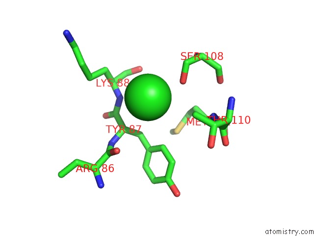
Mono view
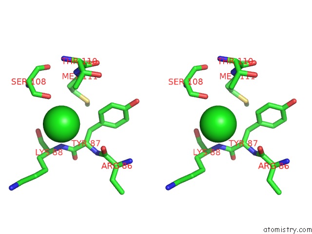
Stereo pair view
Mono view
Stereo pair view
A full contact list of Chlorine with other atoms in the Cl binding
site number 1 of Crystal Structure of A De Novo Designed Protein Homodimer with Curved Beta-Sheet within 5.0Å range:
|
Chlorine binding site 2 out of 2 in 5tph
Go back to
Chlorine binding site 2 out
of 2 in the Crystal Structure of A De Novo Designed Protein Homodimer with Curved Beta-Sheet
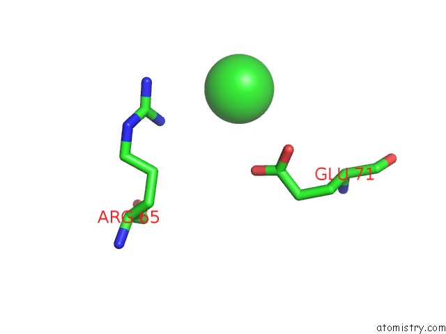
Mono view
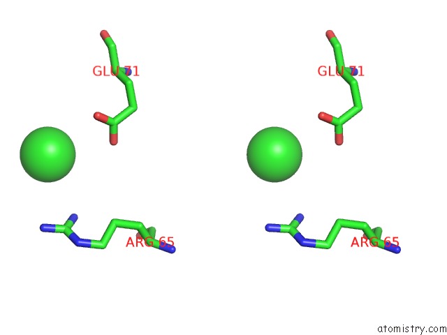
Stereo pair view
Mono view
Stereo pair view
A full contact list of Chlorine with other atoms in the Cl binding
site number 2 of Crystal Structure of A De Novo Designed Protein Homodimer with Curved Beta-Sheet within 5.0Å range:
|
Reference:
E.Marcos,
B.Basanta,
T.M.Chidyausiku,
Y.Tang,
G.Oberdorfer,
G.Liu,
G.V.Swapna,
R.Guan,
D.A.Silva,
J.Dou,
J.H.Pereira,
R.Xiao,
B.Sankaran,
P.H.Zwart,
G.T.Montelione,
D.Baker.
Principles For Designing Proteins with Cavities Formed By Curved Beta Sheets. Science V. 355 201 2017.
ISSN: ESSN 1095-9203
PubMed: 28082595
DOI: 10.1126/SCIENCE.AAH7389
Page generated: Fri Jul 26 17:35:37 2024
ISSN: ESSN 1095-9203
PubMed: 28082595
DOI: 10.1126/SCIENCE.AAH7389
Last articles
Zn in 9J0NZn in 9J0O
Zn in 9J0P
Zn in 9FJX
Zn in 9EKB
Zn in 9C0F
Zn in 9CAH
Zn in 9CH0
Zn in 9CH3
Zn in 9CH1