Chlorine »
PDB 5ti4-5tr9 »
5tpu »
Chlorine in PDB 5tpu: X-Ray Structure of the Wlarb Tdp-Quinovose 3,4-Ketoisomerase From Campylobacter Jejuni
Protein crystallography data
The structure of X-Ray Structure of the Wlarb Tdp-Quinovose 3,4-Ketoisomerase From Campylobacter Jejuni, PDB code: 5tpu
was solved by
H.M.Holden,
J.B.Thoden,
J.Z.Li,
A.S.Riegert,
M.-F.Goneau,
A.M.Cunningham,
E.Vinogradov,
I.C.Schoenhofen,
M.Gilbert,
with X-Ray Crystallography technique. A brief refinement statistics is given in the table below:
| Resolution Low / High (Å) | 30.00 / 2.00 |
| Space group | P 61 |
| Cell size a, b, c (Å), α, β, γ (°) | 104.600, 104.600, 93.900, 90.00, 90.00, 120.00 |
| R / Rfree (%) | 17.9 / 24.5 |
Chlorine Binding Sites:
The binding sites of Chlorine atom in the X-Ray Structure of the Wlarb Tdp-Quinovose 3,4-Ketoisomerase From Campylobacter Jejuni
(pdb code 5tpu). This binding sites where shown within
5.0 Angstroms radius around Chlorine atom.
In total 2 binding sites of Chlorine where determined in the X-Ray Structure of the Wlarb Tdp-Quinovose 3,4-Ketoisomerase From Campylobacter Jejuni, PDB code: 5tpu:
Jump to Chlorine binding site number: 1; 2;
In total 2 binding sites of Chlorine where determined in the X-Ray Structure of the Wlarb Tdp-Quinovose 3,4-Ketoisomerase From Campylobacter Jejuni, PDB code: 5tpu:
Jump to Chlorine binding site number: 1; 2;
Chlorine binding site 1 out of 2 in 5tpu
Go back to
Chlorine binding site 1 out
of 2 in the X-Ray Structure of the Wlarb Tdp-Quinovose 3,4-Ketoisomerase From Campylobacter Jejuni
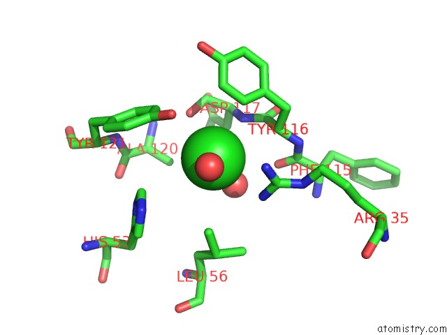
Mono view
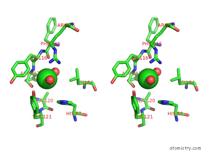
Stereo pair view
Mono view
Stereo pair view
A full contact list of Chlorine with other atoms in the Cl binding
site number 1 of X-Ray Structure of the Wlarb Tdp-Quinovose 3,4-Ketoisomerase From Campylobacter Jejuni within 5.0Å range:
|
Chlorine binding site 2 out of 2 in 5tpu
Go back to
Chlorine binding site 2 out
of 2 in the X-Ray Structure of the Wlarb Tdp-Quinovose 3,4-Ketoisomerase From Campylobacter Jejuni
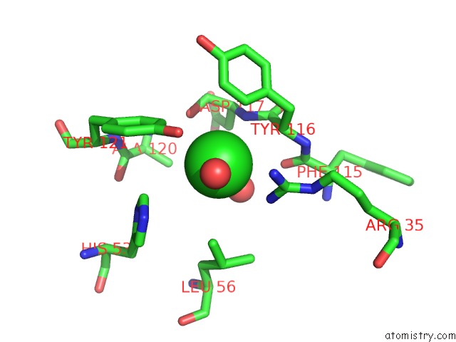
Mono view
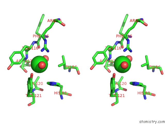
Stereo pair view
Mono view
Stereo pair view
A full contact list of Chlorine with other atoms in the Cl binding
site number 2 of X-Ray Structure of the Wlarb Tdp-Quinovose 3,4-Ketoisomerase From Campylobacter Jejuni within 5.0Å range:
|
Reference:
Z.Z.Li,
A.S.Riegert,
M.F.Goneau,
A.M.Cunningham,
E.Vinogradov,
J.Li,
I.C.Schoenhofen,
J.B.Thoden,
H.M.Holden,
M.Gilbert.
Characterization of the Dtdp-FUC3N and Dtdp-QUI3N Biosynthetic Pathways in Campylobacter Jejuni 81116. Glycobiology V. 27 358 2017.
ISSN: ESSN 1460-2423
PubMed: 28096310
DOI: 10.1093/GLYCOB/CWW136
Page generated: Fri Jul 26 17:36:05 2024
ISSN: ESSN 1460-2423
PubMed: 28096310
DOI: 10.1093/GLYCOB/CWW136
Last articles
Zn in 9J0NZn in 9J0O
Zn in 9J0P
Zn in 9FJX
Zn in 9EKB
Zn in 9C0F
Zn in 9CAH
Zn in 9CH0
Zn in 9CH3
Zn in 9CH1