Chlorine »
PDB 5trc-5u09 »
5ts2 »
Chlorine in PDB 5ts2: Crystal Structure of A Phosphopantetheine Adenylyltransferase (Coad, Ppat) From Pseudomonas Aeruginosa Bound to Dephospho Coenzyme A
Enzymatic activity of Crystal Structure of A Phosphopantetheine Adenylyltransferase (Coad, Ppat) From Pseudomonas Aeruginosa Bound to Dephospho Coenzyme A
All present enzymatic activity of Crystal Structure of A Phosphopantetheine Adenylyltransferase (Coad, Ppat) From Pseudomonas Aeruginosa Bound to Dephospho Coenzyme A:
2.7.7.3;
2.7.7.3;
Protein crystallography data
The structure of Crystal Structure of A Phosphopantetheine Adenylyltransferase (Coad, Ppat) From Pseudomonas Aeruginosa Bound to Dephospho Coenzyme A, PDB code: 5ts2
was solved by
Seattle Structural Genomics Center For Infectious Disease (Ssgcid),
with X-Ray Crystallography technique. A brief refinement statistics is given in the table below:
| Resolution Low / High (Å) | 46.87 / 2.30 |
| Space group | P 21 21 21 |
| Cell size a, b, c (Å), α, β, γ (°) | 98.380, 101.350, 105.710, 90.00, 90.00, 90.00 |
| R / Rfree (%) | 18 / 24.4 |
Other elements in 5ts2:
The structure of Crystal Structure of A Phosphopantetheine Adenylyltransferase (Coad, Ppat) From Pseudomonas Aeruginosa Bound to Dephospho Coenzyme A also contains other interesting chemical elements:
| Calcium | (Ca) | 2 atoms |
Chlorine Binding Sites:
Pages:
>>> Page 1 <<< Page 2, Binding sites: 11 - 11;Binding sites:
The binding sites of Chlorine atom in the Crystal Structure of A Phosphopantetheine Adenylyltransferase (Coad, Ppat) From Pseudomonas Aeruginosa Bound to Dephospho Coenzyme A (pdb code 5ts2). This binding sites where shown within 5.0 Angstroms radius around Chlorine atom.In total 11 binding sites of Chlorine where determined in the Crystal Structure of A Phosphopantetheine Adenylyltransferase (Coad, Ppat) From Pseudomonas Aeruginosa Bound to Dephospho Coenzyme A, PDB code: 5ts2:
Jump to Chlorine binding site number: 1; 2; 3; 4; 5; 6; 7; 8; 9; 10;
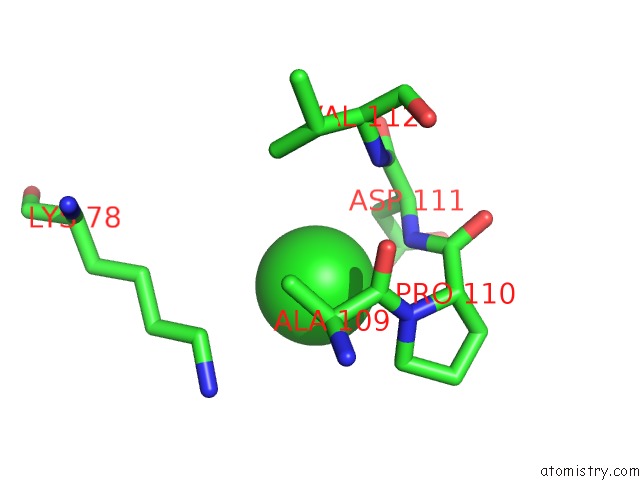
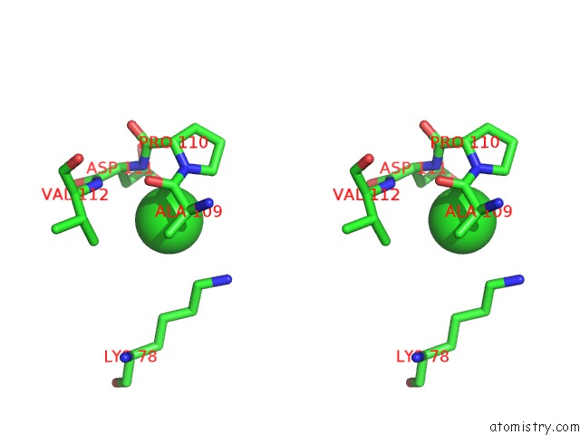
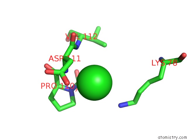
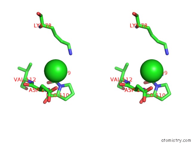
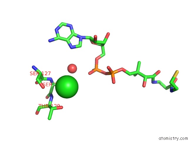
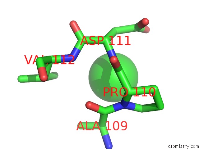
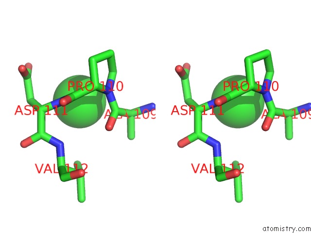
Chlorine binding site 1 out of 11 in 5ts2
Go back to
Chlorine binding site 1 out
of 11 in the Crystal Structure of A Phosphopantetheine Adenylyltransferase (Coad, Ppat) From Pseudomonas Aeruginosa Bound to Dephospho Coenzyme A
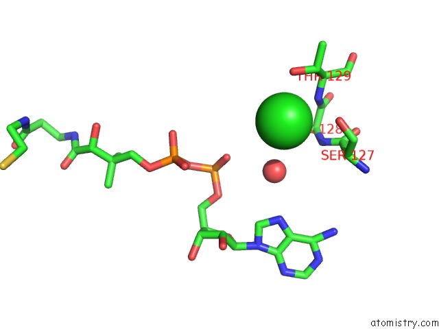
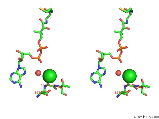
Mono view
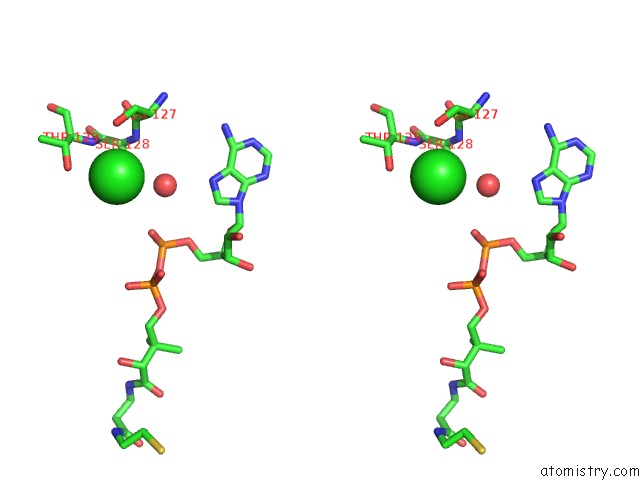
Stereo pair view
Mono view
Stereo pair view
A full contact list of Chlorine with other atoms in the Cl binding
site number 1 of Crystal Structure of A Phosphopantetheine Adenylyltransferase (Coad, Ppat) From Pseudomonas Aeruginosa Bound to Dephospho Coenzyme A within 5.0Å range:
|
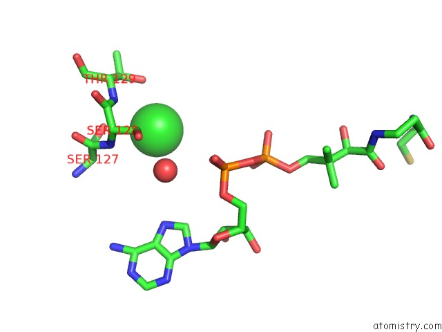
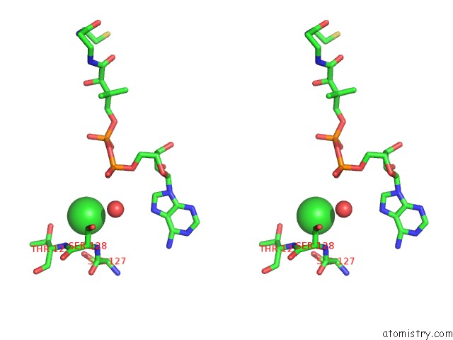
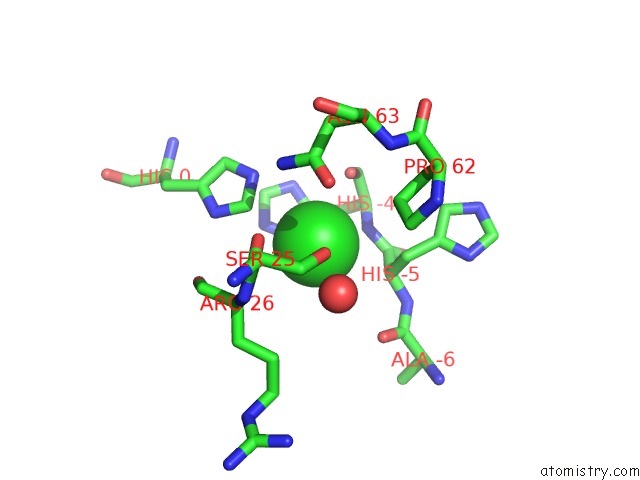
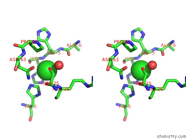
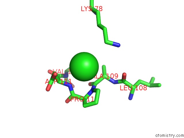
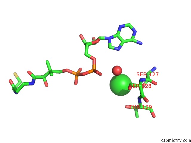
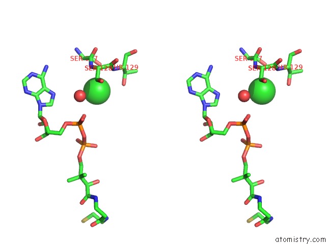
Chlorine binding site 2 out of 11 in 5ts2
Go back to
Chlorine binding site 2 out
of 11 in the Crystal Structure of A Phosphopantetheine Adenylyltransferase (Coad, Ppat) From Pseudomonas Aeruginosa Bound to Dephospho Coenzyme A
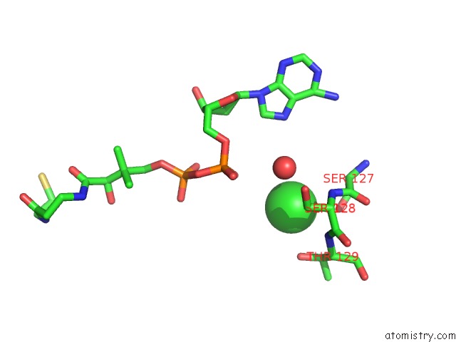
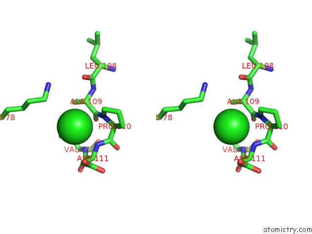
Mono view
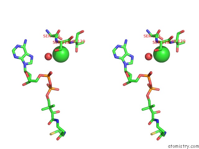
Stereo pair view
Mono view
Stereo pair view
A full contact list of Chlorine with other atoms in the Cl binding
site number 2 of Crystal Structure of A Phosphopantetheine Adenylyltransferase (Coad, Ppat) From Pseudomonas Aeruginosa Bound to Dephospho Coenzyme A within 5.0Å range:
|
Chlorine binding site 3 out of 11 in 5ts2
Go back to
Chlorine binding site 3 out
of 11 in the Crystal Structure of A Phosphopantetheine Adenylyltransferase (Coad, Ppat) From Pseudomonas Aeruginosa Bound to Dephospho Coenzyme A
Mono view
Stereo pair view
Mono view
Stereo pair view
A full contact list of Chlorine with other atoms in the Cl binding
site number 3 of Crystal Structure of A Phosphopantetheine Adenylyltransferase (Coad, Ppat) From Pseudomonas Aeruginosa Bound to Dephospho Coenzyme A within 5.0Å range:
|
Chlorine binding site 4 out of 11 in 5ts2
Go back to
Chlorine binding site 4 out
of 11 in the Crystal Structure of A Phosphopantetheine Adenylyltransferase (Coad, Ppat) From Pseudomonas Aeruginosa Bound to Dephospho Coenzyme A
Mono view
Stereo pair view
Mono view
Stereo pair view
A full contact list of Chlorine with other atoms in the Cl binding
site number 4 of Crystal Structure of A Phosphopantetheine Adenylyltransferase (Coad, Ppat) From Pseudomonas Aeruginosa Bound to Dephospho Coenzyme A within 5.0Å range:
|
Chlorine binding site 5 out of 11 in 5ts2
Go back to
Chlorine binding site 5 out
of 11 in the Crystal Structure of A Phosphopantetheine Adenylyltransferase (Coad, Ppat) From Pseudomonas Aeruginosa Bound to Dephospho Coenzyme A
Mono view
Stereo pair view
Mono view
Stereo pair view
A full contact list of Chlorine with other atoms in the Cl binding
site number 5 of Crystal Structure of A Phosphopantetheine Adenylyltransferase (Coad, Ppat) From Pseudomonas Aeruginosa Bound to Dephospho Coenzyme A within 5.0Å range:
|
Chlorine binding site 6 out of 11 in 5ts2
Go back to
Chlorine binding site 6 out
of 11 in the Crystal Structure of A Phosphopantetheine Adenylyltransferase (Coad, Ppat) From Pseudomonas Aeruginosa Bound to Dephospho Coenzyme A
Mono view
Stereo pair view
Mono view
Stereo pair view
A full contact list of Chlorine with other atoms in the Cl binding
site number 6 of Crystal Structure of A Phosphopantetheine Adenylyltransferase (Coad, Ppat) From Pseudomonas Aeruginosa Bound to Dephospho Coenzyme A within 5.0Å range:
|
Chlorine binding site 7 out of 11 in 5ts2
Go back to
Chlorine binding site 7 out
of 11 in the Crystal Structure of A Phosphopantetheine Adenylyltransferase (Coad, Ppat) From Pseudomonas Aeruginosa Bound to Dephospho Coenzyme A
Mono view
Stereo pair view
Mono view
Stereo pair view
A full contact list of Chlorine with other atoms in the Cl binding
site number 7 of Crystal Structure of A Phosphopantetheine Adenylyltransferase (Coad, Ppat) From Pseudomonas Aeruginosa Bound to Dephospho Coenzyme A within 5.0Å range:
|
Chlorine binding site 8 out of 11 in 5ts2
Go back to
Chlorine binding site 8 out
of 11 in the Crystal Structure of A Phosphopantetheine Adenylyltransferase (Coad, Ppat) From Pseudomonas Aeruginosa Bound to Dephospho Coenzyme A
Mono view
Stereo pair view
Mono view
Stereo pair view
A full contact list of Chlorine with other atoms in the Cl binding
site number 8 of Crystal Structure of A Phosphopantetheine Adenylyltransferase (Coad, Ppat) From Pseudomonas Aeruginosa Bound to Dephospho Coenzyme A within 5.0Å range:
|
Chlorine binding site 9 out of 11 in 5ts2
Go back to
Chlorine binding site 9 out
of 11 in the Crystal Structure of A Phosphopantetheine Adenylyltransferase (Coad, Ppat) From Pseudomonas Aeruginosa Bound to Dephospho Coenzyme A
Mono view
Stereo pair view
Mono view
Stereo pair view
A full contact list of Chlorine with other atoms in the Cl binding
site number 9 of Crystal Structure of A Phosphopantetheine Adenylyltransferase (Coad, Ppat) From Pseudomonas Aeruginosa Bound to Dephospho Coenzyme A within 5.0Å range:
|
Chlorine binding site 10 out of 11 in 5ts2
Go back to
Chlorine binding site 10 out
of 11 in the Crystal Structure of A Phosphopantetheine Adenylyltransferase (Coad, Ppat) From Pseudomonas Aeruginosa Bound to Dephospho Coenzyme A
Mono view
Stereo pair view
Mono view
Stereo pair view
A full contact list of Chlorine with other atoms in the Cl binding
site number 10 of Crystal Structure of A Phosphopantetheine Adenylyltransferase (Coad, Ppat) From Pseudomonas Aeruginosa Bound to Dephospho Coenzyme A within 5.0Å range:
|
Reference:
T.E.Edwards,
D.R.Davies,
J.W.Fairman,
D.Lorimer,
Seattle Structural Genomics Center For Infectious Disease(Ssgcid).
Crystal Structure of A Phosphopantetheine Adenylyltransferase (Coad, Ppat) From Pseudomonas Aeruginosa Bound to Dephospho Coenzyme A To Be Published.
Page generated: Sat Jul 12 09:05:11 2025
Last articles
Fe in 2YXOFe in 2YRS
Fe in 2YXC
Fe in 2YNM
Fe in 2YVJ
Fe in 2YP1
Fe in 2YU2
Fe in 2YU1
Fe in 2YQB
Fe in 2YOO